
Abstract
OBJECTIVE: Technical difficulty during video-assisted thoracoscopic surgery (VATS) lobectomy may not be fully captured by conversion to thoracotomy. We evaluated whether a simple preoperative computed tomography (CT) difficulty score could identify patients at increased risk of difficult VATS lobectomy.
MATERIAL AND METHODS: This single-center retrospective study included patients who underwent planned standard VATS lobectomy with complete CT score data. The author-developed exploratory 0–3 score assigned one point each to the hilar, pleuroparenchymal inflammatory, and obstructive/emphysematous domains. The primary composite endpoint was defined as unplanned conversion to thoracotomy, major intraoperative bleeding, or an operative duration of at least 360 minutes. The duration criterion applied only to completed VATS procedures. The major technical endpoint of the secondary objective was conversion or major bleeding. Scores of 0–1 and 2–3 were defined as low and high CT burden, respectively.
RESULTS: Fifty patients were analyzed; 42 completed VATS and 8 underwent conversion. Major bleeding occurred in two patients, and 11 of 42 completed VATS procedures met the prolonged-duration criterion. The primary and secondary endpoints occurred in 19 (38.0%) and 9 (18.0%) patients, respectively. A high CT burden was present in 12 patients and was associated with higher rates of the primary endpoint (66.7% vs. 28.9%; P = 0.038) and of the secondary endpoint (41.7% vs. 10.5%; P = 0.027). High CT burden was associated with the primary endpoint [odds ratios (ORs) 4.91, 95% confidence interval (CI) 1.22–19.71] and the secondary endpoint (OR 6.07, 95% CI 1.29–28.49). The corresponding area under the curve were 0.634 and 0.672.
CONCLUSION: In this retrospective cohort, a CT difficulty score ≥2 was associated with higher observed rates of difficult VATS and objective major technical events. Discrimination was modest, and confidence intervals were wide; the score is hypothesis-generating rather than validated, and it requires standardized CT reassessment and external validation.
Keywords: Video-assisted thoracoscopic surgery, lobectomy, computed tomography, conversion to thoracotomy, intraoperative bleeding, risk score
Main Points
• An author-developed three-domain preoperative computed tomography (CT) difficulty score classified 12 of the 50 patients as having a high CT burden (score 2–3).
• High CT burden was associated with higher rates of the primary difficult-video-assisted thoracoscopic surgery endpoint (66.7% vs. 28.9%) and the objective major technical endpoint (41.7% vs. 10.5%).
• Event counts were small, and discrimination was modest. The score is exploratory and requires standardized CT reassessment and external validation before clinical use.
INTRODUCTION
Video-assisted thoracoscopic surgery (VATS) lobectomy is a standard minimally invasive approach for appropriately selected patients requiring anatomical lung resection. Randomized trials have shown that VATS can improve early recovery and reduce postoperative pain compared with open thoracotomy without compromising the intended oncological resection in suitable patients., Nevertheless, operations planned through a thoracoscopic approach vary markedly in technical complexity. Dense pleural adhesions, inflammatory or calcified hilar lymph nodes, loss of tissue planes around the pulmonary artery, central obstructive change, and fragile emphysematous parenchyma may transform an otherwise routine lobectomy into a prolonged or hazardous dissection.-
Unplanned conversion to thoracotomy is the most visible and frequently studied manifestation of technical difficulty. A systematic review reported that conversion occurs in approximately one in ten VATS anatomical lung resections, although rates vary with case selection and institutional experience. A recent meta-analysis identified enlarged hilar or mediastinal lymph nodes, pleural adhesions, previous tuberculosis, larger tumors, upper-lobe location, and more complex resections among factors associated with conversion. Conversion alone, however, captures only one end of the spectrum of technical difficulty. Therefore, for the purposes of the present exploratory study, major intraoperative bleeding and markedly prolonged surgery were also included as manifestations of technical difficulty when defining the composite endpoint.
Preoperative CT is useful for technical risk stratification because it directly depicts the hilar, pleural, and parenchymal conditions encountered during surgery. CT features associated with prior granulomatous or inflammatory disease have been linked to conversion. A focused investigation showed that enlarged or calcified lymph nodes and loss of separability from the pulmonary artery may indicate adherent hilar nodes and a higher risk of vascular difficulty. Other groups have developed broader preoperative difficulty models or conversion scores using pulmonary function, previous pulmonary disease, nodal enlargement or calcification, clinical stage, sex, and previous ipsilateral surgery., These studies support preoperative risk enrichment, whereas a concise, anatomically organized CT score may be easier to interpret during routine surgical planning.
The objective of this study was to explore the association between a simple CT-based difficulty score and technical difficulty during planned standard VATS lobectomy. We evaluated both a broad composite endpoint incorporating conversion, major intraoperative bleeding, and prolonged operative duration and a narrower endpoint restricted to objective major technical measures. Because the available cohort was small, the analysis was explicitly exploratory and the manuscript was structured according to relevant TRIPOD reporting principles.
MATERIAL AND METHODS
Study Design and Setting
This single-center retrospective cohort study evaluated lobectomy procedures recorded at a tertiary thoracic surgery unit in Türkiye between April 1, 2022, and June 18, 2026. The institutional database was screened for procedures planned via VATS. According to the study’s inclusion criteria, the operative indication for all included patients was lung cancer. Operations planned from the outset as thoracotomies were ineligible. The study was conceived as an exploratory analysis of score association and discrimination rather than as a definitive clinical prediction-model study.
Study Population and Final Analysis Cohort
Patients with lung cancer scheduled for a standard anatomical VATS lobectomy (right upper, right middle, right lower, left upper, or left lower) were potentially eligible. Procedures that began with VATS and were converted to thoracotomy were retained because conversion was an operative outcome rather than an exclusion criterion. Bilobectomy, extended lobectomy, and lobectomy accompanied by an additional wedge resection were excluded to reduce procedural heterogeneity.
Of 81 procedures coded as planned VATS lobectomy, six complex resections were excluded, leaving 75 eligible standard VATS lobectomies. Preoperative CT examinations could not be retrieved for 24 patients, and one additional patient had insufficient CT data to calculate the complete score. The final analysis cohort, therefore, consisted of 50 patients with complete CT score data, including completed VATS cases and conversion cases. All descriptive, comparative, association, and discriminant analyses reported in this study were restricted to these 50 patients ().
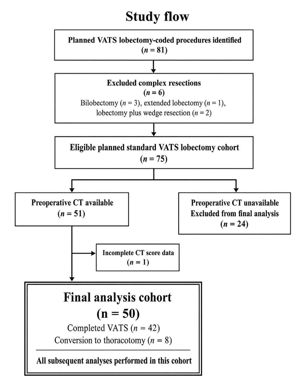
Study flow and formation of the final CT score cohort
VATS: video-assisted thoracoscopic surgery, CT: computed tomography
Outcome Definitions
The primary endpoint, termed difficult VATS, was a composite endpoint defined by the occurrence of at least one of the following: (1) unplanned conversion from a VATS approach to thoracotomy; (2) major intraoperative bleeding; or (3) prolonged operative duration in completed VATS lobectomies. Conversion was counted regardless of its specific technical cause. The duration criterion was applied only to completed VATS procedures because conversion itself already qualified as an event and could independently lengthen the operation.
Operative duration was defined as the skin-to-skin surgical time, from skin incision to completion of skin closure. Any intraoperative frozen-section waiting time between skin incision and skin closure was therefore included in the recorded operative duration. Chest-tube placement performed before skin closure was included; anesthesia induction and emergence were not. Among the 42 completed VATS procedures in the final analysis cohort, the 75th percentile was 359.5 minutes; therefore, an integer duration of at least 360 minutes was defined as a prolonged operation. Major intraoperative bleeding was assigned to the two cases explicitly documented as having blood loss exceeding 500 mL and/or requiring intraoperative erythrocyte-suspension replacement. Exact, continuous measurements of blood loss were unavailable for the remainder of the cohort. A postoperative reoperation for bleeding was not classified as an intraoperative technical event. The major technical endpoint of the secondary objective comprised unplanned conversion or major intraoperative bleeding. Event components were not mutually exclusive.
Preoperative CT Variables and Score Construction
Preoperative CT findings coded in the study database included tumor size and location, chest-wall or pleural contact, pleural thickening or sequelae, pleural calcification, pleural bands or retraction, inflammatory parenchymal sequelae, hilar or interlobar lymph-node short-axis diameter, lymph-node calcification, lymph-node-pulmonary artery contact, emphysema severity, bronchiectasis, and atelectasis or postobstructive change.
The three-domain score was developed by the study authors specifically for this exploratory analysis, using the available preoperative CT variables; it was not a previously published or validated scoring system. To avoid unstable outcome-derived weighting in a small cohort, all domains were weighted equally. The hilar domain contributed one point if at least one of the following was present: hilar or interlobar lymph-node short axis of at least 10 mm, lymph-node calcification, or lymph-node-pulmonary artery contact. The pleuroparenchymal inflammatory domain contributed one point if any of the following were present: pleural thickening or sequelae; pleural band or retraction; inflammatory parenchymal sequelae; or bronchiectasis. The obstructive/emphysematous domain contributed one point when atelectasis, postobstructive change, or moderate-to-severe emphysema was present. Each domain contributed a maximum of one point irrespective of the number of positive findings within that domain, producing a total score from 0 to 3 (). Representative axial CT images corresponding to the three scoring domains are shown in . Fissure completeness was recorded as a separate CT feature when available, but did not contribute a point to the present score. For clinically interpretable exploratory comparisons, scores of 0–1 were categorized as low CT burden, and scores of 2–3 were categorized as high CT burden.
Table 1. Definition of the author-developed exploratory CT difficulty score
Domain | One-point criterion | Rationale |
Hilar | Hilar/interlobar lymph node short axis ≥10 mm, nodal calcification, or lymph node-pulmonary artery contact | Potentially difficult nodal and vascular dissection |
Pleuroparenchymal inflammatory | Pleural thickening/sequel, pleural band/retraction, inflammatory parenchymal sequel, or bronchiectasis | Surrogate of previous pleural or pulmonary inflammation |
Obstructive/emphysematous | Atelectasis/postobstructive change or moderate-to-severe emphysema | Distorted anatomy, impaired exposure, or fragile parenchyma |
Total score | Sum of positive domains | Range 0–3; score ≥2 defined high CT burden |
Each domain contributed a maximum of one point irrespective of the number of positive findings within that domain.
CT: computed tomography
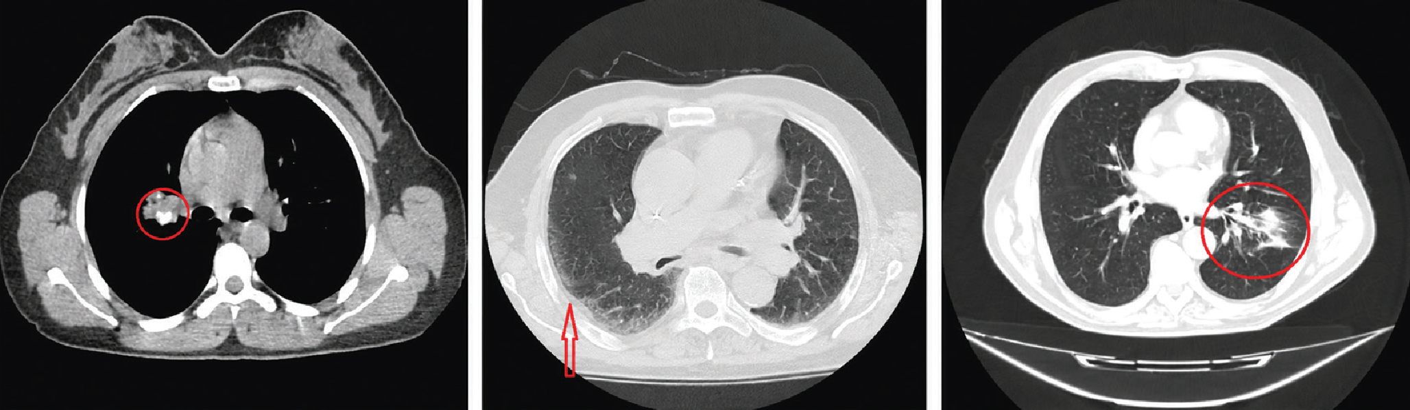
Representative axial computed tomography findings included in the exploratory computed tomography difficulty score. The left image shows a calcified hilar/interlobar lymph node, representing the hilar domain. The middle image shows pleural thickening, representing the pleuroparenchymal inflammatory domain. The right image shows atelectatic or postobstructive parenchymal change, representing the obstructive/emphysematous domain. The presence of at least one qualifying finding within a domain contributed one point, with a maximum of one point per domain
Ethics
The study protocol was approved by the Sakarya University Institutional Ethics Committee (approval no: 578, date: 19.08.2026). Given the retrospective design of the study, the requirement for individual informed consent was waived by the ethics committee.
Statistical Analysis
Continuous variables are presented as median [interquartile range (IQR)], and categorical variables as number (percentage). Baseline characteristics and postoperative outcomes were compared between patients with and without the primary endpoint, using the Mann-Whitney U test for continuous variables and the two-sided Fisher’s exact test for categorical variables. Operative duration was reported descriptively, but not formally compared between primary endpoint groups because prolonged duration was part of the endpoint definition. To assess possible complete-case selection bias, the 24 otherwise eligible patients whose preoperative CT examinations could not be retrieved were compared with the final 50-patient cohort, using the same tests to compare available baseline variables and conversion to thoracotomy. The single patient with a retrievable CT examination, but with incomplete score data, was not included in this comparison.
Operative event rates were compared between the low- and high-burden groups using the two-sided Fisher exact test. No inferential testing was performed across the four score categories because of small cell counts. The association of the score with each endpoint was estimated using univariable logistic regression, first with the score modeled as a one-point increment, and then dichotomized as high versus low CT burden. Results are reported as odds ratios (ORs) with 95% confidence intervals (CIs). Discrimination was assessed using the area under the receiver-operating-characteristic curve (AUC), and 95% CIs were estimated with 5.000 non-parametric bootstrap resamples. Sensitivity, specificity, positive predictive value (PPV), and negative predictive value (NPV) were calculated at a score threshold of at least 2. No random training-test split or multivariable model was used because the final cohort size and event counts were limited. No imputation was performed; analyses were based on available data for each variable. Analyses involving the CT score and study endpoints were restricted to the 50 patients with complete CT score data. Statistical analyses were performed using Python version 3.13.5 (Python Software Foundation, Beaverton, OR, USA). Two-sided P values below 0.05 were considered statistically significant for exploratory inference.
RESULTS
Formation of the Final Analysis Cohort
Of the 81 planned procedures coded as VATS lobectomy, six complex resections were excluded: three bilobectomies, one extended lobectomy, and two lobectomies accompanied by wedge resection. Among the remaining 75 eligible planned standard VATS lobectomies, preoperative CT was unavailable for 24, and the score was incomplete for one. The final analysis cohort comprised 50 patients: 42 underwent VATS and eight required unplanned conversions to thoracotomy. Conversion cases were retained in all analyses ().
Comparison with CT-unavailable Eligible Patients
The 24 otherwise eligible patients who were excluded because their preoperative CT examinations could not be retrieved were similar to the final 50-patient cohort with respect to the measured baseline variables. Conversion occurred in 3 of 24 patients (12.5%) in the CT-unavailable group and in 8 of 50 patients (16.0%) in the final cohort (P = 1.000). No statistically significant differences were detected in age, sex, American Society of Anesthesiologists (ASA) class, smoking status, diabetes mellitus, hypertension, COPD, coronary artery disease, FEV1, laterality, upper-lobe resection, or neoadjuvant treatment (all P > 0.05; ). These measured comparisons do not exclude selection bias from unmeasured factors or those associated with CT availability.
Table 2. Patient characteristics and outcomes in the study cohort and comparison groups
A. Comparison of the final CT-score cohort and otherwise eligible patients excluded because preoperative CT was unavailable | |||||||
|---|---|---|---|---|---|---|---|
Characteristic | Final CT-score cohort (n = 50) | CT unavailable (n = 24) | P value | ||||
Age, years | 66.5 (62.3–72.8) | 67 (59.8–71.2) | 0.623 | ||||
Male sex | 40 (80.0) | 19 (79.2) | 1.000 | ||||
ASA class ≥3 | 37/48 (77.1) | 19/24 (79.2) | 1.000 | ||||
Current/former smoker | 32/49 (65.3) | 14/23 (60.9) | 0.795 | ||||
Diabetes mellitus | 13 (26.0) | 8 (33.3) | 0.586 | ||||
Hypertension | 27 (54.0) | 16 (66.7) | 0.327 | ||||
COPD | 4 (8.0) | 1 (4.2) | 1.000 | ||||
Coronary artery disease | 15 (30.0) | 5 (20.8) | 0.577 | ||||
FEV1, % predicted | 83 (69–92) | 84 (69.5–92) | 0.970 | ||||
Right-sided lobectomy | 29 (58.0) | 17 (70.8) | 0.318 | ||||
Upper-lobe lobectomy | 32 (64.0) | 13 (54.2) | 0.454 | ||||
Neoadjuvant treatment | 5 (10.0) | 3 (12.5) | 0.709 | ||||
Conversion to thoracotomy | 8 (16.0) | 3 (12.5) | 1.000 | ||||
Data are median (interquartile range) or n (%) unless otherwise indicated. Continuous variables were compared using the Mann-Whitney U test and categorical variables using the two-sided Fisher’s exact test. Denominators vary where data were missing. | |||||||
B. Clinical characteristics and postoperative outcomes in the final analysis cohort according to the primary difficult-VATS endpoint | |||||||
Characteristic | Overall (n = 50) | Difficult VATS (n = 19) | Not difficult (n = 31) | P value | |||
Age, years | 66.5 (62.3–72.8) | 65 (62–74.5) | 69 (65–71.5) | 0.833 | |||
Male sex | 40 (80.0) | 16 (84.2) | 24 (77.4) | 0.722 | |||
ASA class ≥3* | 37/48 (77.1) | 14/18 (77.8) | 23/30 (76.7) | 1.000 | |||
Current/former smoker* | 32/49 (65.3) | 13/18 (72.2) | 19/31 (61.3) | 0.541 | |||
Diabetes mellitus | 13 (26.0) | 4 (21.1) | 9 (29.0) | 0.742 | |||
Hypertension | 27 (54.0) | 8 (42.1) | 19 (61.3) | 0.247 | |||
COPD | 4 (8.0) | 1 (5.3) | 3 (9.7) | 1.000 | |||
Coronary artery disease | 15 (30.0) | 5 (26.3) | 10 (32.3) | 0.757 | |||
FEV1, % predicted* | 83 (69–92) | 73 (68–91) | 87 (78–93.8) | 0.103 | |||
Right-sided lobectomy | 29 (58.0) | 9 (47.4) | 20 (64.5) | 0.255 | |||
Upper-lobe lobectomy | 32 (64.0) | 13 (68.4) | 19 (61.3) | 0.764 | |||
Neoadjuvant treatment | 5 (10.0) | 2 (10.5) | 3 (9.7) | 1.000 | |||
Operative duration, min† | 331 (270.5–365) | 377 (361–429) | 299 (226–331) | — | |||
Hospital stay, days | 10 (8–14.8) | 13 (11.5–18.5) | 9 (7–13) | 0.012 | |||
Drain duration, days* | 8 (6–12.5) | 9.5 (7–14.5) | 7 (6–10) | 0.030 | |||
Data are presented as median (interquartile range) or n (%), unless otherwise indicated. Categorical variables were compared using Fisher’s exact test and continuous variables using the Mann-Whitney U test. The primary difficult-VATS endpoint comprised conversion to thoracotomy, major intraoperative bleeding, or prolonged operative duration (≥360 minutes among completed VATS procedures). *Denominators vary because of missing data: ASA class was available in 48 patients, smoking status in 49, FEV1 in 47, and drain duration in 47. Within the difficult-VATS and not-difficult groups, the respective available denominators were 18 and 30 for ASA class, 18 and 31 for smoking status, 17 and 30 for FEV1, and 18 and 29 for drain duration. †Operative duration was not formally compared because prolonged operative duration was a component of the primary endpoint definition. ASA: American Society of Anesthesiologists, FEV1: forced expiratory volume in one second, VATS: video-assisted thoracoscopic surgery, COPD: chronic obstructive pulmonary disease, CT: computed tomography
Clinical Characteristics and Postoperative Outcomes
The median age of the final cohort was 66.5 years (IQR, 62.3–72.8); 40 patients (80.0%) were male. Among patients with available data, ASA class ≥3 was recorded in 37 of 48 patients (77.1%), current or former smoking was recorded in 32 of 49 patients (65.3%), and the median preoperative forced expiratory volume in one second was 83% predicted (IQR, 69–92; n = 47). Twenty-nine lobectomies (58.0%) were right-sided, 32 lobectomies (64.0%) involved an upper lobe, and five patients (10.0%) had received neoadjuvant treatment. All five neoadjuvant-treated patients completed VATS. Two of five patients (40.0%) met the primary difficult-VATS endpoint, both due to prolonged operative durations (380 and 360 minutes); none underwent conversion or experienced major intraoperative bleeding. Their CT scores were 1, 1, 2, 2, and 0. The two event-positive neoadjuvant-treated patients had scores of 2 and 0; the score-2 patient was positive in the pleuroparenchymal inflammatory and obstructive/emphysematous domains.
Nineteen patients met the primary difficult-VATS endpoint, whereas 31 did not. Baseline demographic characteristics, comorbidities, pulmonary function, laterality, lobar location, and neoadjuvant treatment did not differ significantly between the groups. Patients meeting the primary endpoint had longer postoperative hospitalization (median, 13 vs. 9 days; P = 0.012) and longer chest-drain duration (median, 9.5 vs. 7 days; P = 0.030). Operative duration was reported descriptively and was not formally compared between groups because prolonged operative duration was a component of the primary endpoint ().
Operative Events and Endpoint Composition
Unplanned conversion to thoracotomy occurred in eight of 50 patients (16.0%). Major intraoperative bleeding occurred in two patients (4.0%). Among the 42 completed VATS lobectomies, 11 (26.2%) lasted at least 360 minutes. Among the eight conversion cases, the reasons for conversion were adhesions in three patients, marked inflammatory changes involving the fissure in one patient, and massive intraoperative bleeding in one patient. In the remaining three cases, the specific reason for conversion was not separately coded in the database; therefore, no additional cause was assigned retrospectively.
Event components overlapped in two patients: one patient with a CT score of 0 had both major bleeding and prolonged operative duration, and another patient with a CT score of 2 had both major bleeding and conversion. After accounting for these overlaps, the primary composite endpoint occurred in 19 patients (38.0%), and the objective major technical endpoint occurred in nine patients (18.0%) ().
Table 3. Individual operative events according to the exact CT difficulty score
CT difficulty score | Patients, n | Conversion to thoracotomy, n | Major intraoperative bleeding, n | Prolonged operative duration, n/N* | Primary composite endpoint, n | Objective major technical endpoint, n |
|---|---|---|---|---|---|---|
0 | 16 | 1 | 1 | 4/15 | 5 | 2 |
1 | 22 | 2 | 0 | 4/20 | 6 | 2 |
2 | 9 | 4 | 1 | 2/5 | 6 | 4 |
3 | 3 | 1 | 0 | 1/2 | 2 | 1 |
Total | 50 | 8 | 2 | 11/42 | 19 | 9 |
*The numerator is the number of prolonged operations and the denominator is the number of completed VATS procedures within each score category. Event components were not mutually exclusive: one patient with score 0 had both major intraoperative bleeding and prolonged operative duration, and one patient with score 2 had both major intraoperative bleeding and conversion to thoracotomy. No inferential testing was performed across the four individual score categories because of the small cell counts. All analyses were restricted to the final 50-patient cohort. VATS: video-assisted thoracoscopic surgery, CT: computed tomography
CT Score Distribution and Observed Event Rates
The hilar domain was positive in 6 (12.0%) patients, the pleuroparenchymal-inflammatory domain in 25 (50.0%), and the obstructive/emphysematous domain in 18 (36.0%). The total score was 0 in 16 patients, 1 in 22, 2 in 9, and 3 in 3. The primary endpoint occurred in 5 of 16 patients (31.3%) with a score of 0; 6 of 22 (27.3%) with a score of 1; 6 of 9 (66.7%) with a score of 2; and 2 of 3 (66.7%) with a score of 3. The corresponding objective major technical endpoint rates were 2/16 (12.5%), 2/22 (9.1%), 4/9 (44.4%), and 1/3 (33.3%), respectively. Among the 19 patients meeting the primary difficult-VATS endpoint, the hilar, pleuroparenchymal inflammatory, and obstructive/emphysematous domains were positive in 3 (15.8%), 10 (52.6%), and 11 (57.9%), respectively; more than one domain could be positive in the same patient. Because of the small numbers in the individual score strata, these category-specific findings were presented descriptively without inferential testing (, ).

Event rates according to the CT difficulty score. (A) Observed primary difficult-VATS and objective major technical endpoint rates across individual CT difficulty scores; individual score categories are presented descriptively without inferential testing because of small cell counts. (B) Comparison of low CT burden (score 0–1) and high CT burden (score 2–3). ORs, 95% CIs, and P values shown below panel B are from univariable logistic regression
VATS: video-assisted thoracoscopic surgery, CT: computed tomography, CI: confidence interval, OR: odds ratio
Thirty-eight patients had low CT burden (scores 0–1), and 12 had high CT burden (scores 2–3). Conversion was more frequent in the high-burden group than in the low-burden group [5/12 (41.7%) vs. 3/38 (7.9%); P = 0.014]. Major bleeding occurred in 1/12 (8.3%) and in 1/38 (2.6%), respectively (P = 0.426). Prolonged operative duration occurred in 3/7 completed VATS procedures in the high-burden group (42.9%) and in 8/35 completed VATS procedures in the low-burden group (22.9%; P = 0.353). The primary composite endpoint occurred in 8 of 12 high-burden patients (66.7%) compared with 11 of 38 low-burden patients (28.9%; P = 0.038). The objective major technical endpoint occurred in 5 of 12 (41.7%) and 4 of 38 (10.5%), respectively (P = 0.027) (, ).
Table 4. Operative events according to low and high CT difficulty burden in the final analysis cohort
Outcome | Low CT burden (score 0–1, n = 38) | High CT burden (score 2–3, n = 12) | P value |
|---|---|---|---|
Conversion to thoracotomy | 3 (7.9) | 5 (41.7) | 0.014 |
Major intraoperative bleeding | 1 (2.6) | 1 (8.3) | 0.426 |
Prolonged operative duration* | 8/35 (22.9) | 3/7 (42.9) | 0.353 |
Primary composite endpoint† | 11 (28.9) | 8 (66.7) | 0.038 |
Objective major technical endpoint‡ | 4 (10.5) | 5 (41.7) | 0.027 |
Data are n (%) unless otherwise indicated. P values were calculated using the two-sided Fisher’s exact test.
*Prolonged operative duration was defined as ≥360 minutes, corresponding to the upper quartile (75th percentile, 359.5 minutes) among the 42 completed VATS procedures in the final cohort; therefore, the denominator for this row is the number of completed VATS procedures in each CT-burden group.
†The primary composite endpoint comprised conversion to thoracotomy, major intraoperative bleeding, or prolonged operative duration. ‡The objective major technical endpoint comprised conversion to thoracotomy or major intraoperative bleeding.
CT: computed tomography, VATS: video-assisted thoracoscopic surgery
Association and Discrimination of the CT Score
When analyzed as an incremental variable, each one-point increase in the CT score was associated with higher estimated odds of the primary endpoint, although the association did not meet the conventional significance threshold (OR 1.88, 95% CI 0.93–3.81; P = 0.078). The corresponding association with the objective endpoint was similar (OR 2.08, 95% CI 0.90–4.82; P = 0.086).
When dichotomized, high CT burden was associated with the primary difficult-VATS endpoint (OR 4.91, 95% CI 1.22–19.71; P = 0.025) and the objective major technical endpoint (OR 6.07, 95% CI 1.29–28.49; P = 0.022). Discrimination was modest: the AUC was 0.634 (95% bootstrap CI 0.468–0.789) for the primary endpoint and was 0.672 (95% bootstrap CI 0.444–0.881) for the objective endpoint ().
Table 5. Association and discrimination of the CT difficulty score
Performance measure | Primary composite endpoint | Objective major technical endpoint |
|---|---|---|
Events/total | 19/50 (38.0%) | 9/50 (18.0%) |
OR per 1-point increase (95% CI) | 1.88 (0.93–3.81); P = 0.078 | 2.08 (0.90–4.82); P = 0.086 |
AUC (95% bootstrap CI) | 0.634 (0.468–0.789) | 0.672 (0.444–0.881) |
OR for score ≥2 vs. 0–1 (95% CI) | 4.91 (1.22–19.71); P = 0.025 | 6.07 (1.29–28.49); P = 0.022 |
Sensitivity at score ≥2 | 42.1% | 55.6% |
Specificity at score ≥2 | 87.1% | 82.9% |
Positive predictive value | 66.7% | 41.7% |
Negative predictive value | 71.1% | 89.5% |
All analyses were restricted to the final 50-patient cohort. ORs and 95% CIs were estimated using univariable logistic regression. AUC confidence intervals were estimated using 5.000 non-parametric bootstrap resamples. High CT burden was defined as a score ≥2. The primary difficult-VATS endpoint comprised conversion to thoracotomy, major intraoperative bleeding, or prolonged operative duration (≥360 minutes among completed VATS procedures). The objective major technical endpoint comprised conversion to thoracotomy or major intraoperative bleeding.
AUC: area under the curve, CI: confidence interval, CT: computed tomography, OR: odds ratio, VATS: video-assisted thoracoscopic surgery
At a threshold of at least 2 points, sensitivity, specificity, PPV, and NPV for the primary endpoint were 42.1%, 87.1%, 66.7%, and 71.1%, respectively. For the objective endpoint, the corresponding values were 55.6%, 82.9%, 41.7%, and 89.5% ().
DISCUSSION
In this exploratory cohort, a simple three-domain CT score was associated with a higher burden of observed technical events during planned VATS lobectomy. Patients with scores of 2–3 had more conversions and higher rates of both the broad difficult-VATS composite and the narrower objective major technical endpoint than did patients with scores of 0–1. However, the number of events was small, the confidence intervals were wide, and discrimination was modest. The pattern appeared threshold-like rather than strictly linear: scores 0 and 1 had similar event rates, whereas scores 2 and 3 had higher event rates.
The modest AUCs should be interpreted in context, rather than presented as evidence of strong individual-level prediction. The multicenter Epithor conversion score achieved AUCs of approximately 0.62 in development and 0.64 in validation despite being derived from a much larger national database. Our CT score showed a similar range of discrimination, but with substantially greater uncertainty because only 50 patients were analyzed. Conversion itself is clinically heterogeneous: it may be controlled and strategic, or urgent because of bleeding or another major complication. A recent outcome analysis suggests that converted cases should not automatically be equated with failed surgery and may have outcomes comparable with planned thoracotomy when conversion is performed appropriately. A large registry analysis likewise emphasizes that conversion is a relevant operative endpoint but that its impact depends on indication, timing, and patient context.
The broad primary endpoint was intended to capture technical difficulty beyond conversion. In the final cohort, 11 completed VATS procedures were prolonged, and two patients experienced major intraoperative bleeding. The association between high CT burden and the objective endpoint, which excluded operative duration, indicates that the observed signal was not solely generated by the time-based component. Nevertheless, the analysis of bleeding events was underpowered because only two events were documented. A study focused on unexpected VATS bleeding showed that vascular injury and difficult hilar dissection require rapid control strategies and may lead to conversion, transfusion, or extended postoperative recovery. Operative duration is also affected by surgeon experience, team organization, teaching activity, and learning curve; national database data demonstrate that surgical experience can influence conversion and perioperative outcomes. The 360-minute cut-off was an internally derived upper-quartile marker rather than a validated clinical threshold. The relatively long local operative times may reflect differences in institutional workflow, teaching components, case complexity, and surgeon experience; therefore, this threshold should not be applied directly to other centers without recalibration.
The anatomical organization of the score is clinically plausible. The hilar domain reflects enlarged or calcified nodes and loss of a safe tissue plane adjacent to the pulmonary artery. CT-based calcification assessment has previously been associated with conversion risk during VATS lobectomy. The pleuroparenchymal inflammatory domain represents prior inflammation, fibrosis, or pleural reaction that may produce adhesions and obscure normal planes. The obstructive/emphysematous domain represents altered exposure and fragile parenchyma. A published analysis of conversion causes identified adhesions, unexpected nodal fixation, and vascular difficulty as important mechanisms. The present score is deliberately parsimonious and does not encompass every recognized source of VATS difficulty. N1/hilar nodal pathology and more advanced or complex tumor anatomy may materially influence dissection.,,, Neoadjuvant treatment and fissure completeness were not included in the present score and should be explicitly evaluated in larger cohorts. Pleural inflammatory change may primarily increase dissection time in contemporary practice rather than cause a major technical event, but it was retained here as an exploratory marker of altered tissue planes. In the present dataset, lymph-node-pulmonary artery contact was not coded in any patient, and nodal calcification was rare. This likely reduces the sensitivity of the hilar domain and underscores the need for standardized direct image rereading.
The score is more suitable as a planning aid than as a gatekeeper for the surgical approach. A high CT burden could prompt a senior review of the images, explicit preparation to control the proximal pulmonary artery, confirmation of the availability of blood products, and a lower threshold for controlled conversion when safe dissection cannot be maintained. It should not be used to deny VATS because the sensitivity was limited, the high-burden group included only 12 patients, and the confidence intervals were wide. The high NPV for objective major events is potentially useful for reassurance, but is based on only nine events and requires validation.
The statistical interpretation must remain restrained. Prediction tools developed in small datasets can be unstable even when their apparent discrimination is acceptable. PROBAST emphasizes that participant selection, predictor assessment, outcome definition, sample size, missing data, and analysis methods all affect risk of bias and applicability. Formal sample-size guidance for binary-outcome prediction models generally requires substantially more patients and events than were available here. We therefore avoided outcome-derived point weights, did not fit a multivariable model or split the dataset into development and test subsets, and reported the score as exploratory. These choices reduce unnecessary model complexity but do not remove optimism, selection bias, or uncertainty.
Study Limitations
This study has several limitations. First, the study was retrospective and single-center, and only 50 patients had complete CT score data. A comparison with the 24 otherwise eligible patients whose preoperative CT could not be retrieved did not identify statistically significant differences in the measured baseline variables or conversion rate (); however, this cannot exclude selection bias due to unmeasured characteristics or factors related to CT availability. One additional patient had incomplete score data. Second, CT variables were derived from findings historically coded in the database rather than from a standardized, blinded dual-reader reassessment. Reader variability and misclassification may, therefore, affect reproducibility; interobserver agreement could not be measured. Third, the score and pragmatic threshold were evaluated in the same cohort, without external validation or formal calibration assessment. Fourth, exact blood-loss measurements were unavailable for most patients; therefore, classification of major bleeding relied on explicit documentation of blood loss above 500 mL and/or intraoperative replacement with erythrocyte suspension. Fifth, the prolonged-duration threshold was internally derived from the 42 completed VATS procedures and was longer than typical VATS lobectomy durations; it may reflect local workflow, teaching activity, case mix, and surgeon experience, and it should not be considered externally transportable without recalibration. Sixth, surgeon identity, cumulative experience, and detailed operative approach were not modeled. Conversion causes were incompletely coded, with no specific cause recorded in three of the eight conversion cases. Seventh, individual high-score categories were sparse, particularly the score of 3, which contained only three patients. Finally, fissure completeness and the systematically graded difficulty of pleural or hilar dissection were not components of the current score.
Future work should include standardized thin-section CT rereading by at least two blinded observers; explicit grading of the lymph-node-pulmonary artery interface, pleural inflammatory burden, and fissural anatomy; and prospective capture of estimated blood loss, vascular injury, adhesion severity, hilar dissection difficulty, surgeon experience, and conversion cause. A larger, multicenter cohort should then be used to assess calibration, clinical utility, and external transportability of the score.
CONCLUSION
In this small, single-center cohort of 50 patients undergoing planned, standard VATS lobectomy, a preoperative CT difficulty score of at least 2 was associated with higher observed rates of conversion and of composite technical difficulty. Discrimination was modest, event counts were low, and effect estimates were imprecise, with wide confidence intervals. The score should be viewed as a hypothesis-generating framework for standardized CT assessment and operative planning, not as a validated tool for selecting patients for VATS or excluding patients from VATS.
Ethics
Ethics Committee Approval: The study protocol was approved by the Sakarya University Institutional Ethics Committee (approval no: 578, date: 19.08.2026).
Informed Consent: Given the retrospective design of the study, the requirement for individual informed consent was waived by the ethics committee.
Footnotes
Authorship Contributions
Concept: İ.D., R.A., B.O., Design: İ.D., R.A., B.O., Data Collection or Processing: İ.D., R.A., B.O., A.N., Y.A., A.Ş., Analysis or Interpretation: İ.D., R.A., B.O., A.N., Y.A., A.Ş., Literature Search: İ.D., A.N., Y.A., A.Ş., Writing: İ.D., R.A., B.O., A.N., Y.A., A.Ş.
Conflict of Interest
No conflict of interest was declared by the authors.
Financial Disclosure
The authors declared that this study received no financial support.
References
Lim E, Batchelor TJP, Dunning J, et al. Video-assisted thoracoscopic or open lobectomy in early-stage lung cancer. NEJM Evid. 2022;1(3):EVIDoa2100016.
Bendixen M, Jørgensen OD, Kronborg C, Andersen C, Licht PB. Postoperative pain and quality of life after lobectomy via video-assisted thoracoscopic surgery or anterolateral thoracotomy for early stage lung cancer: a randomised controlled trial. Lancet Oncol. 2016;17(6):836-844.
Power AD, Merritt RE, Abdel-Rasoul M, Moffatt-Bruce SD, D’Souza DM, Kneuertz PJ. Estimating the risk of conversion from video-assisted thoracoscopic lung surgery to thoracotomy-a systematic review and meta-analysis. J Thorac Dis. 2021;13(2):812-823.
Wang S, Yan H, Wen J, Zhou Z, Xu J. Risk factors for conversion to thoracotomy in patients with lung cancer undergoing video-assisted thoracoscopic surgery: a meta-analysis. PLoS One. 2024;19(11):e0313236.
Lim CG, Shin KM, Lim JS, et al. Predictors of conversion to thoracotomy during video-assisted thoracoscopic surgery lobectomy in lung cancer: additional predictive value of FDG-PET/CT in a tuberculosis endemic region. J Thorac Dis. 2017;9(8):2427-2436.
Takeda T, Matsuoka S, Miura K, Hamanaka K, Shimizu K, Eguchi T. Prediction of pulmonary artery-adherent lymph nodes for minimally invasive lung resection. Ann Thorac Surg. 2022;114(3):969-977.
Wang Z, Wang Y, Sun D. A retrospective and prospective study to establish a preoperative difficulty predicting model for video-assisted thoracoscopic lobectomy and mediastinal lymph node dissection. BMC Surg. 2022;22(1):135.
Yoo GY, Yoon SK, Moon MH, Moon SW, Hwang W, Kim KS. Development of a risk scoring model to predict unexpected conversion to thoracotomy during video-assisted thoracoscopic surgery for lung cancer. J Chest Surg. 2024;57(3):302-311.
Collins GS, Reitsma JB, Altman DG, Moons KG. Transparent reporting of a multivariable prediction model for individual prognosis or diagnosis (TRIPOD): the TRIPOD statement. BMJ. 2015;350:g7594.
Fourdrain A, Georges O, Gossot D, et al. Patient risk factors for conversion during video-assisted thoracic surgery-the Epithor conversion score. Eur J Cardiothorac Surg. 2022;62(3):ezac249.
Taylor M, Raj Krishna G, Rammohan K, et al. Outcomes after conversion from video-assisted thoracoscopic lobectomy to thoracotomy. Thorac Cardiovasc Surg. 2024;72(5):394-401.
Bongiolatti S, Gonfiotti A, Viggiano D, et al. Risk factors and impact of conversion from VATS to open lobectomy: analysis from a national database. Surg Endosc. 2019;33(12):3953-3962. Erratum in: Surg Endosc. 2019;33(12):3963.
Chen W, Jin K, Yu Z, Liu H. Analysis of risk factors and management of unexpected intraoperative bleeding during video-assisted thoracic surgery for non-small cell lung cancer: a case-control study. J Thorac Dis. 2023;15(5):2729-2741.
Bertolaccini L, Fornaro G, Ciani O, et al. The impact of surgical experience in VATS lobectomy on conversion and patient quality of life: results from a comprehensive national video-assisted thoracic surgical database. Cancers (Basel). 2023;15(2):410.
Samson P, Guitron J, Reed MF, Hanseman DJ, Starnes SL. Predictors of conversion to thoracotomy for video-assisted thoracoscopic lobectomy: a retrospective analysis and the influence of computed tomography-based calcification assessment. J Thorac Cardiovasc Surg. 2013;145(6):1512-1518.
Puri V, Patel A, Majumder K, et al. Intraoperative conversion from video-assisted thoracoscopic surgery lobectomy to open thoracotomy: a study of causes and implications. J Thorac Cardiovasc Surg. 2015;149(1):55-61, 62.e1.
Riley RD, Collins GS. Stability of clinical prediction models developed using statistical or machine learning methods. Biom J. 2023;65(8):e2200302.
Wolff RF, Moons KGM, Riley RD, et al. PROBAST: a tool to assess the risk of bias and applicability of prediction model studies. Ann Intern Med. 2019;170(1):51-58.
Riley RD, Snell KI, Ensor J, et al. Minimum sample size for developing a multivariable prediction model: part II - binary and time-to-event outcomes. Stat Med. 2019;38(7):1276-1296. Erratum in: Stat Med. 2019;38(30):5672.