
Abstract
OBJECTIVE: Chronic obstructive pulmonary disease (COPD) is linked to systemic inflammatory activity and an increased burden of cardiovascular comorbidity, particularly coronary artery disease (CAD). This study aimed to examine serum asprosin, fatty acid-binding protein 4 (FABP4), and vaspin levels in COPD patients according to the presence or absence of CAD.
MATERIAL AND METHODS: This prospective cross-sectional study included 120 participants: COPD (n = 40), COPD with CAD (n = 40), and healthy controls (n = 40). Serum biomarker levels were compared among groups. Receiver operating characteristic analysis and multivariable logistic regression were used to assess discriminative performance and independent associations.
RESULTS: Biomarker levels differed significantly among groups (P < 0.001). Median asprosin levels were 8.32, 14.98, and 11.88 ng/mL; median FABP4 levels were 205.86, 708.63, and 416.18 ng/L; and median vaspin levels were 4.37, 6.17, and 5.50 ng/mL in the COPD, COPD+CAD, and control groups, respectively. Area under the curve values were 0.898 for asprosin, 0.891 for vaspin, and 0.860 for FABP4 (all P < 0.001). After adjustment for age and low-density lipoprotein (LDL) cholesterol, asprosin [odds ratio (OR) 1.965, 95% confidence interval (CI) 1.436–2.691], FABP4 (OR 1.014, 95% CI 1.007–1.021), and vaspin (OR 2.896, 95% CI 1.767–4.745) remained independently associated with CAD (all P < 0.001).
CONCLUSION: Serum asprosin, FABP4, and vaspin levels were elevated in COPD patients with CAD, and showed discriminative ability in this cohort, with each biomarker remaining associated with CAD after adjustment for age and LDL cholesterol. These findings require confirmation in larger prospective cohorts.
Keywords: Asprosin, COPD, coronary artery disease, FABP4, vaspin
Main Points
• Serum asprosin, fatty acid-binding protein 4 (FABP4), and vaspin concentrations were lowest in patients with chronic obstructive pulmonary disease (COPD) alone, intermediate in healthy controls, and highest in patients with COPD and coronary artery disease (CAD).
• Among patients with COPD, asprosin and vaspin had numerically higher area under the curve values for CAD than those for FABP4.
• Each biomarker remained associated with CAD in a separate multivariable model adjusted for age and low-density lipoprotein cholesterol.
• The receiver operating characteristic-derived cut-off values are exploratory and require validation before clinical use.
INTRODUCTION
Chronic obstructive pulmonary disease (COPD) is a respiratory condition in which persistent airflow limitation is accompanied by ongoing inflammatory changes in the airways and lung parenchyma. Its clinical impact extends beyond respiratory symptoms: COPD contributes substantially to global morbidity and mortality, and often coexists with extrapulmonary conditions. Systemic inflammation, oxidative stress, and endothelial dysfunction have been implicated as possible pathways linking COPD with increased cardiovascular risk.
Cardiovascular diseases, particularly coronary artery disease (CAD), are major contributors to mortality in patients with COPD. Previous studies suggest that COPD is associated with a higher risk of CAD, even after accounting for shared risk factors such as smoking and age. Chronic hypoxemia, systemic inflammation, and vascular dysfunction are considered important contributors to this relationship. Patients with both COPD and CAD generally have poorer clinical outcomes, including higher rates of hospitalization, exacerbations, and mortality. Therefore, biomarkers that may help characterize cardiovascular involvement in COPD have become a focus of increasing clinical interest.
Adipokine-related pathways have gained increasing attention in cardiometabolic research, particularly adipokines such as asprosin, fatty acid-binding protein 4 (FABP4), and vaspin.- Asprosin, a fasting-induced glucogenic protein released from adipose tissue, has been reported to contribute to endothelial dysfunction and vascular inflammation. Elevated serum asprosin concentrations have been described in patients with atherosclerosis, and experimental evidence has implicated asprosin in myocardial infarction-related pathways., FABP4, an adipocyte-derived lipid chaperone, is involved in macrophage activation and vascular inflammation, and higher circulating concentrations have been linked to increased cardiovascular risk and coronary plaque instability. Similarly, vaspin, a serine protease inhibitor derived from visceral adipose tissue, has been reported to contribute to anti-inflammatory regulation and improved insulin sensitivity, although its precise cardiovascular role remains incompletely understood. Despite these findings, data on the role of these markers in COPD-related CAD remain limited and inconsistent, highlighting the need for targeted research in this area.
The present study evaluated serum asprosin, FABP4, and vaspin levels in patients with COPD, stratified by the presence of CAD, and examined their association with CAD within the COPD cohort. Biomarker profiles were also compared with those of healthy controls to explore whether these markers may be related to cardiovascular comorbidity in COPD.
MATERIAL AND METHODS
Study Design and Ethics Approval
This prospective cross-sectional study was conducted at a tertiary care hospital from May to August 2025. Ethical approval was obtained from the Ethics Committee of Atatürk University Faculty of Medicine (approval no: B.30.2.ATA.0.01.00/426, date: 30.05.2025). All study procedures followed the principles of the Declaration of Helsinki, and written informed consent was provided by each participant before enrollment.
Study Population
A total of 120 participants were enrolled and categorized into three groups: 40 patients with stable COPD; 40 patients with COPD and angiographically or scintigraphically confirmed CAD; and 40 healthy control subjects matched for age, sex, and body mass index (BMI). None of the participants had an established or documented CAD diagnosis at enrollment. All patients with COPD underwent a cardiology evaluation as part of the study protocol. Following this assessment, patients with suspected CAD underwent invasive coronary angiography or stress myocardial perfusion imaging as clinically indicated. All CAD cases were newly diagnosed, and blood samples were collected during the diagnostic evaluation before the initiation of lipid-lowering therapy. Only clinically stable patients with CAD were included, whereas individuals with acute coronary syndromes were excluded.
COPD was defined according to the Global Initiative for Chronic Obstructive Lung Disease 2025 criteria, based on a post-bronchodilator FEV1/FVC ratio <0.70. All COPD patients were former smokers who had stopped active smoking at least 12 months before enrollment. Exclusion criteria included recent exacerbation (<8 weeks), active infection, systemic inflammatory or autoimmune disease, malignancy, chronic kidney or liver disease, diabetes mellitus, and systemic corticosteroid use. Control subjects were non-smokers with no chronic diseases and no current medication use.
Sample Size Calculation
The sample size was calculated before enrollment using G*Power version 3.1.9.7 (Heinrich Heine University, Düsseldorf, Germany). The calculation was based on a Kruskal-Wallis test comparing three independent groups (COPD, COPD+CAD, and healthy controls), with an alpha error probability of 0.05 and a statistical power of 80% (1–β = 0.80). The effect size was determined from previously published serum vaspin data reported by Stavileci and Koldaş, corresponding to a medium effect (Cohen’s f ≈ 0.30). This approach yielded a minimum sample size of 111 participants, equivalent to 37 individuals per group. To allow for possible missing data or protocol deviations, the target sample size was set at 120 participants, with 40 participants in each group.
Definition of Coronary Artery Disease
CAD was defined in accordance with the current European Society of Cardiology guidelines. Patients were considered to have CAD if invasive coronary angiography demonstrated ≥50% luminal stenosis in at least one major coronary artery or if stress perfusion imaging showed ≥10% myocardial ischemia.
Clinical and Functional Assessments
All participants underwent a comprehensive clinical evaluation, including a detailed medical history, smoking status (pack-years), and BMI. Symptom severity was evaluated with the COPD Assessment Test (CAT) and the modified Medical Research Council (mMRC) dyspnea scale. Pulmonary function testing was performed according to the 2019 ATS/ERS technical standards. A trained technician conducted measurements using a body plethysmography device (Cosmed Q-Box, Rome, Italy). Participants were instructed to avoid smoking during the preceding 24 hours, heavy meals during the preceding 2 hours, and vigorous physical activity during the 30 minutes before testing. Calibration was performed daily, and measurements were corrected to body temperature and pressure-saturated conditions. The best of three acceptable post-bronchodilator maneuvers was used in the analysis.
Echocardiographic Evaluation
Transthoracic echocardiography was performed on all participants by the same cardiologist using a Toshiba S270-A echocardiography system fitted with a 2.5 MHz transducer. The procedure was conducted in the left lateral decubitus position under resting conditions. Cardiac structures and function were evaluated using two-dimensional and M-mode imaging together with color, pulsed-wave, and continuous-wave Doppler. Mean pulmonary artery pressure (mPAP) was estimated from the tricuspid regurgitation jet velocity combined with an assessment of right atrial pressure.
Blood Sampling and Biochemical Analysis
For biochemical testing, venous blood was drawn from each participant. After collection, the tubes were kept upright at room temperature for 10–20 minutes to allow clot formation and were then centrifuged at 4000 rpm for 15 minutes at +4 °C. Serum fractions were separated into aliquots and kept in a −80°C freezer until the day of the assay.
Determination of Analytes
To avoid inter-assay variation, serum asprosin, vaspin, and FABP4 levels were measured on the same day. Concentrations were determined by enzyme-linked immunosorbent assay (ELISA) using commercial kits: Human Asprosin ELISA kit (Cat. No: E4095Hu), Human Vaspin ELISA kit (Cat. No: E0921Hu), and Human FABP4 ELISA kit (Cat. No: E7430Hu), all from BTLAB (China), following the manufacturer’s protocol.
The detection ranges for asprosin, vaspin, and FABP4 kits were 0.5–100 ng/mL, 0.05–10 ng/mL, and 20–7000 ng/L, respectively. Assay precision was indicated by intra-assay and inter-assay coefficients of variation of <8% and <10%, respectively.
ELISA Procedure
96-well microplates pre-coated with monoclonal antibodies specific for human asprosin, vaspin, or FABP4 were used. Serum samples and serially diluted standards were added to the wells, allowing the target molecules to bind to the capture antibodies. Unbound substances were washed away. A biotin-conjugated detection antibody specific to each analyte was added, followed by a second wash and an incubation with streptavidin-horseradish peroxidase. The enzymatic reaction was initiated by adding the substrate solution, 3,3’,5,5’-tetramethylbenzidine, resulting in a color change proportional to the concentration of the target analyte. After the stop solution had been added, optical density was measured at 450 nm using a microplate reader. Analyte concentrations were derived from the absorbance-concentration standard curve generated from known standards.
Statistical Analysis
Data were analyzed with SPSS version 27.0 (IBM Corp., Armonk, NY, USA) and MedCalc Statistical Software version 22.0 (MedCalc Software Ltd, Ostend, Belgium). The Kolmogorov-Smirnov test was used to evaluate the distribution of continuous variables. Variables were expressed as mean±standard deviation or median (interquartile range), depending on data distribution. Group comparisons were performed using Student’s t-test for normally distributed variables and the Mann-Whitney U test for non-normally distributed variables. To identify factors associated with CAD among COPD patients, a univariate logistic regression analysis was first performed. Asprosin, FABP4, and vaspin were subsequently evaluated in separate multivariable logistic regression models, each adjusted for age and low-density lipoprotein (LDL) cholesterol. Age and LDL cholesterol were included as clinically relevant potential confounders because they differed between the COPD and COPD+CAD groups. Separate models were constructed for each biomarker to maintain model parsimony given the number of CAD events. Results were reported as odds ratios (OR) with 95% confidence intervals (CI). The number of variables included in each multivariable model was limited to reduce the risk of model overfitting.
The discriminative performance of asprosin, vaspin, and FABP4 for CAD among COPD patients was evaluated using receiver operating characteristic (ROC) curve analysis. Area under the curve (AUC) values with 95% confidence intervals were calculated. Cut-off values were determined according to the Youden index, and corresponding sensitivity and specificity values were reported. The ROC-derived cut-off values were considered exploratory because they were both derived and evaluated within the same cohort without internal or external validation. A P value <0.05 was considered statistically significant.
RESULTS
A total of 120 participants were included in the analysis: 40 patients with COPD, 40 patients with both COPD and CAD (COPD+CAD), and 40 healthy controls. The median age was 64.0 years (58.0–68.0) in the COPD group and 71.0 years (60.0–77.25) in the COPD+CAD group (P = 0.04). Sex distribution was comparable across the three groups (P > 0.05). The median BMI was 27.0 kg/m (23.0–33.0) in the COPD group and 27.5 kg/m (24.75–31.25) in the COPD+CAD group (P = 0.96). The control group was chosen to be broadly comparable with respect to age, sex, and BMI.
Pulmonary function parameters were similar in the COPD and COPD+CAD groups. Forced expiratory volume in one second (FEV1) (L) values were 1.72 (1.11–2.28) and 1.645 (1.10–2.33) (P = 0.83), while FEV1 (%) values were 71.0 (53.0–86.0) and 65.0 (48.25–88.25), respectively (P = 0.81). FVC (L) was 3.10 (1.99–3.85) in the COPD group and 2.935 (1.9575–3.5725) in the COPD+CAD group (P = 0.63); FVC (%) was 93.0 (73.0–109.0) and 95.5 (67.5–106.25), respectively (P = 0.81). Cigarette consumption in pack-years was also comparable [30.0 (20.0–40.0) vs. 29.0 (20.0–45.75), P = 0.58]. Symptom scores did not differ statistically between groups: CAT was 9.0 (5.0–10.0) in the COPD group and 10.0 (7.0–15.0) in the COPD+CAD group (P = 0.06), while mMRC was 1.0 (1.0–2.0) in the COPD group and 2.0 (1.0–3.0) in the COPD+CAD group (P = 0.08). Hospitalization counts [0 (0–0) vs. 0 (0–1), P = 0.35] and COPD exacerbation counts [0 (0–0) vs. 0 (0–1), P = 0.39] were also comparable between groups ().
Table 1. Comparison of demographic characteristics, pulmonary function parameters, and laboratory findings between the COPD and COPD+CAD groups
| COPD group n = 40 Median (IQR) | COPD+CAD group n = 40 Median (IQR) | P |
|---|---|---|---|
Age (year) | 64.0 (58.0–68.0) | 71.0 (60.0–77.25) | 0.04 |
BMI (kg/m2) | 27.0 (23.0–33.0) | 27.5 (24.75–31.25) | 0.96 |
FEV1 (L) | 1.72 (1.11–2.28) | 1.645 (1.10–2.33) | 0.83 |
FEV1 (%) | 71.0 (53.0–86.0) | 65.0 (48.25–88.25) | 0.81 |
FVC (L) | 3.10 (1.99–3.85) | 2.935 (1.9575–3.5725) | 0.63 |
FVC (%) | 93.0 (73.0–109.0) | 95.5 (67.5–106.25) | 0.81 |
Cigarette (pack/year) | 30.0 (20.0–40.0) | 29.0 (20.0–45.75) | 0.58 |
CAT | 9.0 (5.0–10.0) | 10.0 (7.0–15.0) | 0.06 |
mMRC | 1.0 (1.0–2.0) | 2.0 (1.0–3.0) | 0.08 |
Hospitalization | 0 (0–0) | 0 (0–1) | 0.35 |
COPD exacerbation* | 0 (0–0) | 0 (0–1) | 0.39 |
Triglyceride (mg/dL) | 99.0 (70.0–125.0) | 109.5 (75.0–175.5) | 0.45 |
Total cholesterol (mg/dL) | 170.0 (138.25–188.5) | 187.0 (155.0–216.0) | 0.02 |
LDL (mg/dL) | 94.5 (69.675–107.0) | 102.0 (89.0–130.0) | 0.02 |
HDL (mg/dL) | 46.0 (35.0–62.0) | 41.5 (33.75–51.5) | 0.18 |
CRP (mg/L) | 5.0 (3.0–9.0) | 6.2 (3.4725–10.375) | 0.21 |
WBC (cells/µL) | 8350 (6620–10330) | 8070 (6342.5–10297.5) | 0.78 |
Hemoglobin (g/dL) | 15.0 (14.1–16.2) | 15.45 (14.05–16.925) | 0.48 |
Hematocrit (%) | 45.7 (44.0–48.1) | 46.2 (40.85–51.35) | 0.59 |
Lymphocyte (cells/µL) | 2170 (1530–3010) | 2115 (1575–2597.5) | 0.73 |
Eosinophil (cells/µL) | 140 (100–280) | 185 (87.5–390) | 0.67 |
Neutrophil (cells/µL) | 5330 (4020–6390) | 5025 (3567.5–7095) | 0.62 |
Platelet (cells/µL) | 274000 (230000–332000) | 257000 (193750–319000) | 0.42 |
mPAP (mmHg) | 30.0 (28.0–35.0) | 35.0 (30.0–46.25) | 0.06 |
*COPD exacerbation: number of COPD exacerbations in the last year. COPD: chronic obstructive pulmonary disease, CAD: coronary artery disease, FEV1: volume of air expelled in the 1st second of forced expiration, FVC: forced vital capacity, mPAP: mean pulmonary artery pressure, BMI: body mass index, CAT: COPD assessment test, mMRC: modified Medical Research Council, IQR: interquartile range, LDL: low-density lipoprotein, HDL: high-density lipoprotein, CRP: C-reactive protein, WBC: white blood cell count
Among laboratory variables, total cholesterol and LDL cholesterol levels differed significantly between groups. Total cholesterol was 170.0 (138.25–188.5) in the COPD group and 187.0 (155.0–216.0) in the COPD+CAD group (P = 0.02), and LDL cholesterol was 94.5 (69.675–107.0) in the COPD group and 102.0 (89.0–130.0) in the COPD+CAD group (P = 0.02). Triglyceride and HDL cholesterol levels were not significantly different (P = 0.45 and P = 0.18, respectively). CRP, WBC, hemoglobin, hematocrit, lymphocyte, eosinophil, neutrophil, and platelet counts were comparable between groups (P > 0.05 for all). mPAP was 30.0 (28.0–35.0) in the COPD group and 35.0 (30.0–46.25) in the COPD+CAD group (P = 0.06) ().
Significant differences in serum biomarker levels were observed among the three groups (all P < 0.001). Asprosin levels were 8.32 (7.42–10.35) ng/mL in the COPD group, 14.98 (13.97–16.48) ng/mL in the COPD+CAD group, and 11.88 (9.68–13.21) ng/mL in the control group. Similarly, FABP4 levels were 205.86 (166.80–258.31) ng/L in the COPD group, 708.63 (521.30–820.48) ng/L in the COPD+CAD group, and 416.18 (275.47–623.18) ng/L in the control group. Vaspin levels were 4.37 (3.45–4.55) ng/mL in the COPD group, 6.17 (5.86–7.08) ng/mL in the COPD+CAD group, and 5.50 (3.69–6.36) ng/mL in the control group ().
Table 2. Comparison of serum asprosin, FABP4, and vaspin levels among the COPD, COPD+CAD, and control groups
| COPD group n = 40 Median (IQR) | COPD+CAD group n = 40 Median (IQR) | Control n = 40 Median (IQR) | P |
Asprosin (ng/mL) | 8.32 (7.42–10.35) | 14.98 (13.97–16.48) | 11.88 (9.68–13.21) | <0.001 |
FABP4 (ng/L) | 205.86 (166.80–258.31) | 708.63 (521.30–820.48) | 416.18 (275.47–623.18) | <0.001 |
Vaspin (ng/mL) | 4.37 (3.45–4.55) | 6.17 (5.86–7.08) | 5.50 (3.69–6.36) | <0.001 |
COPD: chronic obstructive pulmonary disease, CAD: coronary artery disease, FABP4: fatty acid-binding protein 4, IQR: interquartile range
ROC curve analyses are presented in . Asprosin and vaspin showed discriminative performance in detecting the presence of CAD in patients with COPD (AUC = 0.898 and 0.891, respectively; both P < 0.001), while FABP4 showed comparatively lower discriminative performance (AUC = 0.860, P < 0.001) in this cohort. The Youden index-derived cut-off values were >11.27 ng/mL for asprosin, >252.88 ng/L for FABP4, and >4.69 ng/mL for vaspin. At these thresholds, asprosin showed 87% sensitivity and 72% specificity, FABP4 showed 90% sensitivity and 72% specificity, and vaspin showed 87% sensitivity and 80% specificity. These cut-off values were derived from the present sample and lack external validation; therefore, caution is warranted in their interpretation.
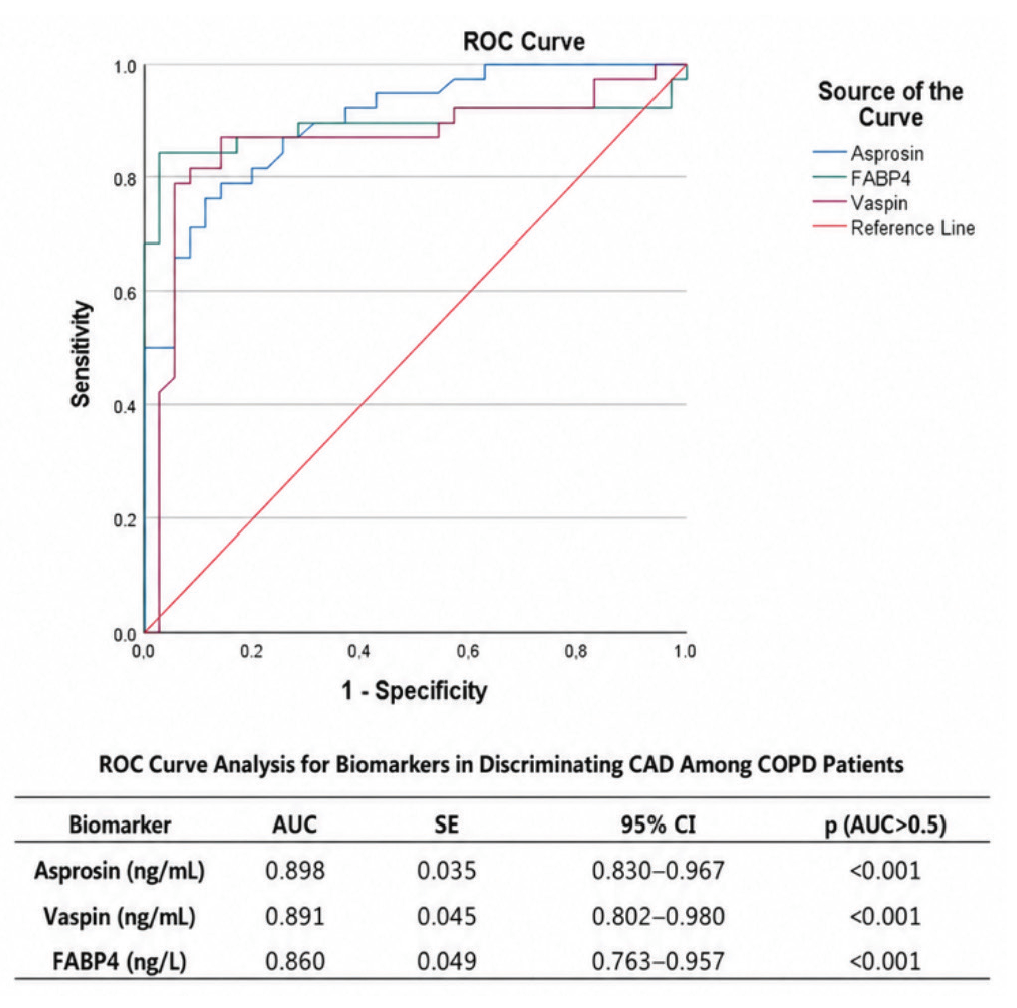
ROC curves evaluating the discriminative performance of serum asprosin, FABP4, and vaspin for the presence of coronary artery disease in patients with chronic obstructive pulmonary disease
FABP4: fatty acid-binding protein 4, ROC: receiver operating characteristic, CI: confidence interval, AUC: area under the curve, COPD: chronic obstructive pulmonary disease, CAD: coronary artery disease
Univariable and multivariable logistic regression analyses are presented in . In univariable analyses, higher serum levels of asprosin (OR 1.928, 95% CI 1.447–2.568; P < 0.001), FABP4 (OR 1.010, 95% CI 1.005–1.014; P < 0.001), and vaspin (OR 2.859, 95% CI 1.777–4.601; P < 0.001) were associated with the presence of CAD among patients with COPD. LDL cholesterol and total cholesterol were also associated with CAD in univariable analyses. In separate multivariable models adjusted for age and LDL cholesterol, all three biomarkers remained independently associated with CAD. The adjusted ORs were 1.965 (95% CI 1.436–2.691; P < 0.001) for asprosin, 1.014 (95% CI 1.007–1.021; P < 0.001) for FABP4, and 2.896 (95% CI 1.767–4.745; P < 0.001) for vaspin.
Table 3. Univariable and multivariable logistic regression analyses for the presence of CAD in patients with COPD
Variable | Univariable OR (95% CI) | P | Adjusted OR (95% CI) | P |
|---|---|---|---|---|
Age (year) | 1.047 (0.999–1.096) | 0.054 | — | — |
LDL (mg/dL) | 1.023 (1.004–1.042) | 0.017 | — | — |
Total cholesterol (mg/dL) | 1.016 (1.003–1.030) | 0.019 | — | — |
HDL (mg/dL) | 0.968 (0.935–1.002) | 0.065 | — | — |
Triglyceride (mg/dL) | 1.004 (0.996–1.012) | 0.336 | — | — |
BMI (kg/m2) | 0.989 (0.908–1.077) | 0.797 | — | — |
COPD exacerbation* | 1.158 (0.699–1.919) | 0.568 | — | — |
Number of hospitalizations | 1.725 (0.678–4.390) | 0.252 | — | — |
Asprosin (ng/mL) | 1.928 (1.447–2.568) | <0.001 | 1.965 (1.436–2.691)† | <0.001 |
FABP4 (ng/L) | 1.010 (1.005–1.014) | <0.001 | 1.014 (1.007–1.021)‡ | <0.001 |
Vaspin (ng/mL) | 2.859 (1.777–4.601) | <0.001 | 2.896 (1.767–4.745)§ | <0.001 |
*Number of COPD exacerbations in the last year. †Model 1: adjusted for age and LDL cholesterol. ‡Model 2: adjusted for age and LDL cholesterol. §Model 3: adjusted for age and LDL cholesterol. COPD: chronic obstructive pulmonary disease, CAD: coronary artery disease, OR: odds ratio, CI: confidence interval, BMI: body mass index, LDL: low-density lipoprotein, HDL: high-density lipoprotein, FABP4: fatty acid-binding protein 4
DISCUSSION
In this study, which evaluated adipokine profiles in relation to CAD status in patients with COPD, we observed that serum levels of asprosin, FABP4, and vaspin were lower in COPD patients than in healthy controls. Conversely, patients with both COPD and CAD exhibited significantly higher levels of all three biomarkers than those in the control and COPD-only groups. Among these markers, asprosin and vaspin had higher AUC values than FABP4 for distinguishing COPD patients with CAD from those without. Furthermore, in separate multivariable models adjusted for age and LDL cholesterol, asprosin, FABP4, and vaspin each remained independently associated with the presence of CAD.
COPD is characterized by persistent airflow limitation and chronic pulmonary inflammation, as well as extrapulmonary manifestations affecting the metabolic and cardiovascular systems. Previous studies have indicated that certain adipokines, such as FABP4 and vaspin, may be dysregulated in patients with chronic lung diseases. FABP4 levels have been reported to be reduced in the airways of individuals with COPD, while vaspin levels appear to decrease in response to pulmonary inflammation and lung injury. However, to date, no studies have directly evaluated serum asprosin concentrations in COPD patients.-
Chronic airway inflammation in COPD is associated with sustained activation of innate immune pathways that may influence adipokine signaling at both local and systemic levels. In the setting of chronic airway inflammation, altered macrophage function within the COPD airway may modify adipokine expression patterns, potentially shifting the balance between pro-inflammatory and regulatory mediators. Disruption of adipokine signaling has been linked to amplification of inflammatory cascades, including NF-κB–mediated pathways, which may extend beyond the pulmonary compartment. Through these mechanisms, chronic airway inflammation may contribute to endothelial dysfunction and vascular inflammation, providing a plausible biological link between chronic airway disease and increased cardiovascular vulnerability. Nevertheless, the association between circulating adipokines and cardiovascular disease in this population remains insufficiently defined. Given that COPD is considered an independent risk factor for CAD, the interplay between adipokine dysregulation and cardiovascular involvement warrants further attention.
Circulating levels of asprosin, FABP4, and vaspin have been reported to be elevated in CAD.,- Asprosin has been associated with metabolic and vascular alterations relevant to atherogenesis, and a possible relationship between circulating asprosin and regional adiposity has also been reported. Higher serum asprosin levels have been reported in CAD patients than in healthy individuals. FABP4 has similarly been associated with cardiovascular and metabolic disorders, with increased levels reported in CAD and metabolic syndrome. Vaspin levels have also been reported to be elevated in CAD, particularly in those with metabolic comorbidities., Collectively, these findings suggest that these adipokines may be associated with cardiometabolic alterations relevant to CAD; however, the underlying mechanisms cannot be established in the present study.
In our study, asprosin, FABP4, and vaspin demonstrated discriminative ability for CAD in patients with COPD. In addition, all three biomarkers remained independently associated with the presence of CAD in separate multivariable models, each adjusted for age and LDL cholesterol. The consistency between the univariate and adjusted analyses suggests that the observed associations were not explained solely by differences in age or LDL cholesterol between the COPD groups. However, these findings should be interpreted in the context of the study’s cross-sectional design and relatively small sample size. The ROC-derived cut-off values were obtained from the same cohort and were neither internally nor externally validated; therefore, their clinical applicability cannot be established from the present study. These adipokines were significantly associated with CAD among COPD patients, whereas most clinical and functional parameters were not. This suggests that certain adipokines may be related to cardiovascular comorbidity in COPD. Moreover, the observation that serum levels of these biomarkers were higher not only in patients with COPD and comorbid CAD but also in healthy controls, compared to those with isolated COPD, may reflect differential regulation related to the burden of both cardiovascular and pulmonary disease. Furthermore, the finding that biomarker levels in the COPD-only group were lower than in healthy controls and were highest in those with coexisting CAD suggests that this pattern does not follow a simple linear inflammatory gradient. Although BMI was comparable between COPD groups, it does not adequately reflect body composition or adipose tissue distribution. COPD is frequently associated with regional alterations in fat mass, reductions in fat-free mass, and sarcopenic phenotypes. In addition, chronic systemic inflammation and oxidative stress may contribute to functional changes in adipose tissue, potentially influencing adipokine secretion independently of total body weight. Therefore, the observed biomarker distribution may reflect heterogeneity in metabolic regulation rather than a monotonic progression of systemic inflammation. The distribution pattern of biomarkers across the study groups may support the hypothesis that systemic metabolic and inflammatory pathways are associated with cardiovascular pathology in COPD. Importantly, asprosin, FABP4, and vaspin remained independently associated with CAD after adjustment for age and LDL cholesterol. However, given the cross-sectional design and lack of validation, these findings should be interpreted as associations rather than as evidence of predictive utility, and require confirmation in prospective studies.
Study Limitations
This study has certain limitations. Its cross-sectional design precludes causal inference, and reverse causation cannot be excluded. Although all patients with COPD underwent cardiology evaluation, further testing for CAD was performed only when clinically indicated. Silent or subclinical CAD may therefore not have been fully excluded in the COPD-only group, potentially introducing verification bias. The relatively limited sample size may restrict generalizability, although strict inclusion criteria were applied to ensure group comparability. It also precluded age-stratified analyses of potential age-related differences in biomarker associations. Although BMI was comparable between COPD groups, direct measures of body composition, including fat mass and fat distribution, were not performed, preventing assessment of their influence on circulating biomarker levels. Temporal changes in biomarker levels could not be assessed. The absence of external validation may also limit the generalizability of the discriminative findings. The very high AUC values should be interpreted with caution because they may be overestimated in this modest sample size. Although the associations remained significant after adjustment for age and LDL cholesterol, residual confounding by unmeasured cardiovascular and metabolic factors cannot be excluded.
Nevertheless, several aspects of the study design may support the interpretation of our findings. The inclusion of three clearly defined groups, including healthy controls selected to be comparable in age, sex, and BMI, allowed direct comparisons of biomarkers. CAD was identified during the study, and only clinically stable patients were included before treatment initiation, thereby potentially reducing treatment-related confounding. Strict exclusion criteria for diabetes mellitus, active inflammatory conditions, malignancy, and systemic corticosteroid use may have further reduced confounding of adipokine levels. Biomarker measurements were performed under standardized conditions, and both ROC and multivariable analyses allowed the assessment of discriminative performance and independent associations.
CONCLUSION
Our findings suggest that serum levels of asprosin, FABP4, and vaspin are associated with CAD among patients with COPD in this cohort. Since CAD was identified during study-related cardiovascular evaluation, these adipokines may reflect underlying cardiovascular involvement rather than represent established diagnostic markers. The observed biomarker gradients among healthy individuals, individuals with isolated COPD, and individuals with COPD and CAD may reflect overlapping inflammatory and metabolic pathways associated with cardiovascular comorbidity. Although further prospective longitudinal studies are required to validate these results, our findings contribute to the understanding of possible links between adipokine regulation and cardiovascular disease in COPD.
Ethics
Ethics Committee Approval: Ethical approval was obtained from the Ethics Committee of Atatürk University Faculty of Medicine (approval no: B.30.2.ATA.0.01.00/426, date: 30.05.2025).
Informed Consent: Written informed consent was obtained from all participants prior to study enrollment.
Footnotes
Authorship Contributions
Surgical and Medical Practices: A.A., B.K., M.B.C., B.N.G., Concept: A.A., Design: A.A., L.S., Data Collection or Processing: A.A., B.K., M.B.C., B.N.G., E.L., Analysis or Interpretation: A.A., B.K., M.B.C., E.L., L.S., Literature Search: B.N.G., Writing: A.A., B.K., M.B.C., B.N.G., L.S.
Conflict of Interest
No conflict of interest was declared by the authors.
Financial Disclosure
The authors declared that this study received no financial support.
Declaration Regarding the Use of AI and AI-Assisted Technologies: During the preparation of this manuscript, the authors used ChatGPT (OpenAI) solely to assist with editing language, improving clarity, and organizing the manuscript. The tool was not used for data generation, statistical analysis, or scientific decision-making. All AI-assisted content was critically reviewed and revised by the authors, who take full responsibility for the final manuscript.
References
Kostikas K, Hillas G, Gogali A. 2025 GOLD report: what is new and what is noteworthy for the practicing clinician. Copd. 2025;22(1):2451613.
Agusti A, Vogelmeier CF. GOLD 2024: a brief overview of key changes. J Bras Pneumol. 2023;49(6):e20230369.
Li XF, Wan CQ, Mao YM. Analysis of pathogenesis and drug treatment of chronic obstructive pulmonary disease complicated with cardiovascular disease. Front Med (Lausanne). 2022;9:979959.
Brassington K, Selemidis S, Bozinovski S, Vlahos R. Chronic obstructive pulmonary disease and atherosclerosis: common mechanisms and novel therapeutics. Clin Sci (Lond). 2022;136(6):405-423.
Wang G, Fan C, Chai Y, et al. Association of serum asprosin concentrations with heart failure. BMC Cardiovascular Disorders. 2023;23(1):617.
Zhang Z, Zhu L, Wang Z, Hua N, Hu S, Chen Y. Can the new adipokine asprosin be a metabolic troublemaker for cardiovascular diseases? A state-of-the-art review. Prog Lipid Res. 2023;91:101240.
Hung WC, Yu TH, Wu CC, et al. FABP3, FABP4, and heart rate variability among patients with chronic schizophrenia. Front Endocrinol (Lausanne). 2023;14:1165621.
Kobat MA, Celik A, Balin M, et al. The investigation of serum vaspin level in atherosclerotic coronary artery disease. J Clin Med Res. 2012;4(2):110-113.
Mazur-Bialy AI. Asprosin - a fasting-induced, glucogenic, and orexigenic adipokine as a new promising player. Will it be a new factor in the treatment of obesity, diabetes, or infertility? A review of the literature. Nutrients. 2021;13(2):620.
Zhang Z, Tan Y, Zhu L, et al. Asprosin improves the survival of mesenchymal stromal cells in myocardial infarction by inhibiting apoptosis via the activated ERK1/2-SOD2 pathway. Life Sci. 2019;231:116554.
Furuhashi M. Fatty acid-binding protein 4 in cardiovascular and metabolic diseases. J Atheroscler Thromb. 2019;26(3):216-232.
Kerget B, Kerget F, Kahraman ÇY, Aksakal A, Araz Ö. The relationship between NLRP3 rs10159239 and vaspin rs2236242 gene variants and obstructive sleep apnea. Ups J Med Sci. 2021:126.
Li Q, Chen R, Moriya J, et al. A novel adipocytokine, visceral adipose tissue-derived serine protease inhibitor (vaspin), and obesity. J Int Med Res. 2008;36(4):625-629.
Stavileci B, Koldaş ZL. The relationship between vaspin, nesfatin-1 plasma levels and presence of fragmented QRS with the severity of coronary atherosclerosis. Adv Med Sci. 2022;67(2):298-303.
Zhang SJ, Qin XZ, Zhou J, et al. Adipocyte dysfunction promotes lung inflammation and aberrant repair: a potential target of COPD. Front Endocrinol (Lausanne). 2023;14:1204744.
Perea L, Rodrigo-Troyano A, Cantó E, et al. Reduced airway levels of fatty-acid binding protein 4 in COPD: relationship with airway infection and disease severity. Respir Res. 2020;21(1):21.
Aslani MR, Ghazaei Z, Ghobadi H. Correlation of serum fatty acid binding protein-4 and interleukin-6 with airflowlimitation and quality of life in stable and acute exacerbation of COPD. Turk J Med Sci. 2020;50(2):337-345.
Wang C. Obesity, inflammation, and lung injury (OILI): the good. Mediators Inflamm. 2014;2014(1):978463.
Smekal A, Vaclavik J. Adipokines and cardiovascular disease: a comprehensive review. Biomed Pap Med Fac Univ Palacky Olomouc Czech Repub. 2017;161(1):31-40.
Yigitdol I, Gulumsek E, Demirtas D, et al. The role of serum asprosin levels in predicting the severity of coronary artery disease in patients with diabetes mellitus. Ir J Med Sci. 2024;193(3):1239-1247.
Moradi N, Fouani FZ, Vatannejad A, Bakhti Arani A, Shahrzad S, Fadaei R. Serum levels of asprosin in patients diagnosed with coronary artery disease (CAD): a case-control study. Lipids Health Dis. 2021;20(1):88.
Jalilian N, Pakzad R, Shahbazi M, et al. Circulating FABP-4 levels in patients with atherosclerosis or coronary artery disease: a comprehensive systematic review and meta-analysis. Cardiovasc Ther. 2023;2023(1):1092263.
Aksakal A, Kerget B, Özkan HB, Laloğlu E, Araz Ö, Akgün M. Serum asprosin levels in obstructive sleep apnea syndrome: relationship with disease severity and adipose tissue distribution. Sleep Breath. 2026;30(1):18.
Blüher M. Vaspin in obesity and diabetes: pathophysiological and clinical significance. Endocrine. 2012;41(2):176-182.
Benz E, Trajanoska K, Lahousse L, et al. Sarcopenia in COPD: a systematic review and meta-analysis. Eur Respir Rev. 2019;28(154):190049.