
Abstract
OBJECTIVE
Chest wall reconstruction following oncologic resection must achieve structural stability while allowing normal respiratory mechanics. This uncontrolled retrospective case series describes our experience with steel wire–based chest wall reconstruction. The findings are intended to demonstrate the feasibility and safety of this approach. Twisted stainless steel wires (No. 5) offer a low-cost, semi-rigid alternative with passive physiological micro-mobility.
MATERIAL AND METHODS
A retrospective case series was conducted in the Department of Thoracic Surgery at Services Hospital in Lahore, Pakistan, from May 2023 to April 2025. Thirty-eight patients who underwent oncologic chest wall resection and reconstruction with twisted stainless steel wires were included.
RESULTS
All 38 patients underwent successful wire-based reconstruction; chondrosarcoma was the most frequent cause (21.1% of cases). Mean operative time was 185±56 minutes, and mean blood loss was 295±120 mL. Postoperative pain decreased rapidly (visual analogue scale 4.1 at 24 h → 2.0 at 48 h → 1.7 by day 7). Chest wall recoil was preserved in all patients, with no paradoxical motion or mechanical instability. Functional recovery was excellent: 79% had normal chest expansion on clinical assessment at 1 month, which increased to 94% by 3 months, with complete resolution of dyspnea. The complication rate was low (seroma 2.6%, chronic pain 2.6%); no infections, hematomas, wire failures, or 30-day mortality occurred.
CONCLUSION
Twisted stainless steel wires provide a safe, semi-rigid, and cost-effective solution for reconstructing oncologic chest wall defects. This technique preserves respiratory mechanics, ensures functional and cosmetic outcomes, and costs less than 40 USD per case in our setting, making it especially valuable for low- and middle-income countries.
Keywords: Chest wall resection, reconstruction, stainless steel wires, polymethyl methacrylate
Main Points
• Twisted stainless steel wire chest wall reconstruction provides effective semi-rigid stabilization after oncologic chest wall resection while preserving physiological chest wall motion.
• The technique demonstrated excellent functional recovery with rapid pain reduction, preserved chest wall recoil, and no mechanical instability.
• Postoperative morbidity was minimal, with no infections, wire failures, or early mortality observed in this series.
• Wire-based reconstruction is highly cost-effective (40 USD), making it a practical alternative to rigid prostheses in low- and middle-income countries.
• This study represents the largest reported Pakistani case series evaluating steel wire–based chest wall reconstruction, addressing a regional evidence gap.
INTRODUCTION
Chest wall resection for tumors creates complex defects that require reconstruction to restore structural integrity, protect intrathoracic organs, and preserve respiratory mechanics. Because the chest wall comprises both rigid bony and flexible soft-tissue components essential for ventilation, reconstruction must ensure stability, maintain physiological motion, minimize pain, and reduce postoperative complications. A wide range of techniques and materials has been described for soft-tissue and skeletal reconstruction., For reconstruction, rigid prosthetics, including titanium plates, synthetic meshes, and polymethyl methacrylate (PMMA) bone cement, have been widely used., Although PMMA provides compressive strength and ease of application, its rigid, non-adhesive nature is associated with thermal necrosis, infection, chronic pain, limited tensile strength, and impaired respiratory dynamics due to rigid reconstruction.- Titanium mesh and plating systems offer improved biocompatibility and radiopacity, and have shown favorable outcomes in high-volume oncologic centers such as Memorial Sloan Kettering.- However, in low-resource settings, the high cost of bone cement, titanium mesh, and rigid plating systems limits their widespread use.
Stainless steel wires have re-emerged as a practical, low-cost alternative for chest wall reconstruction. Twisted No. 5 steel wires provide semi-rigid stability while permitting passive chest wall micro-mobility, thereby maintaining respiratory mechanics better than static cement-based patches. They also reduce postoperative discomfort, allow adaptive mobility under respiratory forces, and remain widely accessible and inexpensive, making them particularly suitable for low- and middle-income countries. Internationally, hybrid techniques using steel wires to reinforce neoribs have demonstrated improved tensile and compressive strength while avoiding complications associated with cement application adjacent to medullary bone., A recent report highlighted the simplicity and effectiveness of non-rigid wire reconstruction for rib chondrosarcoma.
Despite their advantages, evidence on wire-based reconstruction in Pakistan is scarce. Only a single case report, describing successful neo-sternum creation using twisted steel wires for recurrent sternal chondrosarcoma, has been published to date. No observational studies or case series have evaluated this technique regionally, leaving a substantial knowledge gap, particularly in resource-limited healthcare systems where affordable alternatives to rigid prostheses are urgently needed.
This study addresses this gap by reporting our institutional experience with chest wall reconstruction using twisted stainless steel wires. Through subjective evaluation of respiratory function, chest wall recoil, postoperative pain, and complications, we aim to generate evidence that may guide broader adoption of this cost-effective technique and improve outcomes in settings where economic constraints shape surgical decision-making.
MATERIAL AND METHODS
Study Design and Setting
This uncontrolled, retrospective, single-center case series was conducted at the Department of Thoracic Surgery, Services Hospital, Lahore, Pakistan. All consecutive patients who underwent chest wall resection for tumors, followed by reconstruction using No. 5 twisted stainless steel wires between May 2023 and April 2025 were included. The study was registered retrospectively at ClinicalTrials.gov (NCT07148648). Institutional Review Board approval was obtained from the Services Institute of Medical Sciences, Services Hospital, Pakistan (decision no.: IRB/2025/1653/SIMS) on 21.08.2025. This study involved analysis of existing medical records without direct patient contact. Informed consent was waived by the Institutional Review Board. All patient data were anonymized to protect confidentiality, and the study adhered to the Declaration of Helsinki and institutional ethical standards. Written informed consent was obtained from all patients for the publication of their clinical data and any accompanying images.
Study Population
Patients were eligible if they were aged 15 years or older, underwent chest wall resection requiring reconstruction with stainless steel wires, had defects involving ≥3 ribs and/or measuring ≥5 cm in maximum diameter, and reconstruction performed using stainless steel wires with or without mesh reinforcement. Patients were excluded if two or fewer ribs were resected, if the defect was located beneath the scapula or over the liver, or if reconstruction was completed using only mesh and muscle. Defects beneath the scapula involving ribs 2–4 posteriorly were classified as “scapula-adjacent”; in these cases, the scapula provided inherent, rigid protection, and only polypropylene mesh reinforcement, without wire placement, was applied to the resected rib edges. Similarly, defects over the liver do not produce paradoxical movement during respiration due to the ligamentous attachments of the liver; therefore, these defects are reinforced with mesh alone. Additional exclusions included contraindications to major surgery; pre-existing chest wall deformities (scoliosis, pectus excavatum, pectus carinatum); severe respiratory disease, defined as forced expiratory volume in one second <40% predicted; cases requiring reoperation or experiencing intraoperative events that altered the reconstructive plan; and patients with incomplete medical records.
Surgical Technique
All procedures were performed under general anesthesia with single- or double-lumen endotracheal intubation to facilitate lung isolation when required. No. 5 stainless steel wire (0.5 mm) was used for rib fixation, providing an optimal balance between tensile strength and flexibility while minimizing the risk of rib cut-through. The wires were manually contoured and anchored either circumferentially around the rib stumps or through predrilled holes in the rib remnants, depending on bone quality and the surgeon’s preference.
Chest wall reconstruction was performed using a structured two-step wire fixation technique to achieve optimal stability. Initially, vertical wires were placed across the defect by passing a circumferential wire around the rib immediately superior to the defect and then around the rib immediately inferior to the defect. These wires were twisted 6–8 rotations to approximate the rib margins and to restore continuity without excessive tightening. A total of 3–6 vertical wires were applied, depending on defect size.
Subsequently, horizontal wires were placed starting from the posterior rib stump and advanced toward the anterior margin of the defect. These were twisted by 3–5 rotations at each fixation point and interlocked sequentially with the previously placed vertical wires, creating a reinforced lattice configuration. This resulted in a stable interlocking stainless steel wire-mesh construct, providing enhanced rigidity while preserving physiological chest-wall compliance. Following steel-wire reconstruction, a double-layer polypropylene mesh (e.g., Prolene mesh, Ethicon Prolene Mesh) was applied as an onlay reinforcement patch to the reconstructed defect. The mesh was tailored to overlap the defect margins by at least 2–3 cm and secured to surrounding ribs, intercostal muscles, or fascia using interrupted non-absorbable sutures.
Following skeletal stabilization, soft-tissue coverage was achieved using primary closure, local or regional muscle flaps, or rotational flaps. Extensive defects were reconstructed in collaboration with the plastic surgery team when required. Representative operative steps are illustrated in , , . Postoperatively, patients received intravenous broad-spectrum antibiotics for 5–7 days, along with standard analgesia and pulmonary physiotherapy.
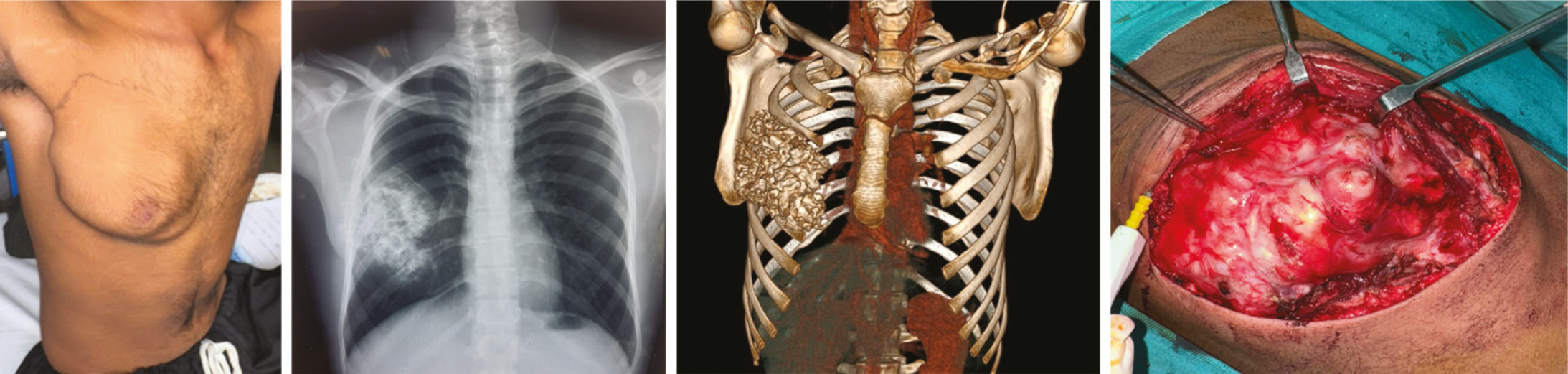
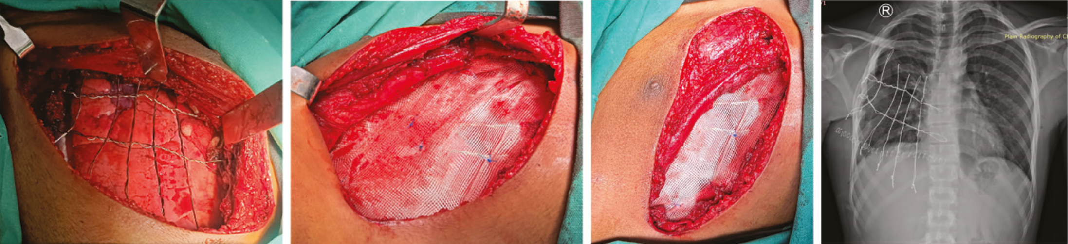

DIEP: deep inferior epigastric perforator
Postoperative Analgesia and Pain Assessment
Postoperative analgesia followed a standardized multimodal protocol of intravenous paracetamol and nonsteroidal anti-inflammatory drugs unless contraindicated, and was supplemented with intravenous morphine (0.05–0.1 mg/kg) as required. Thoracic epidural or paravertebral blockade was used selectively, based on the extent of resection or the anesthetist’s preference. Rescue analgesia was administered when visual analogue scale (VAS) pain scores were ≥4 at rest or ≥5 during movement. Analgesic use was not converted to morphine milligram equivalents; thus, comparisons remain descriptive. Pain assessments were conducted at 24 hours, 48 hours, and 7 days postoperatively.
Chest Wall Movement and Recoil Assessment
Chest wall function was evaluated subjectively and clinically at discharge, at 1-month follow-up, and at 3-month follow-up by the operating surgeon or thoracic fellow. Patients who underwent surgery earlier in the study period remained under active surveillance, providing a total follow-up duration of up to two years. Assessment included inspection for paradoxical movement during quiet and deep breathing and, when feasible, measurement of axillary excursion. Normal chest movement was defined as symmetrical expansion or ≥3 cm axillary excursion. Restricted movement was defined as <3 cm excursion, ≥25% asymmetry relative to the contralateral side. Chest wall recoil was considered intact in the absence of paradoxical inward movement on clinical examination and of a flail segment on postoperative chest radiography.
Data Collection
Data extracted from medical records included demographics, comorbidities, tumor diagnosis, resection characteristics, intraoperative details, and postoperative outcomes. The primary outcomes were postoperative pain (VAS scores) and chest wall mobility. Secondary outcomes included operative time, blood loss, intensive care unit (ICU) stay, hospital stay, postoperative complications, need for reoperation, and 30-day mortality. Follow-up duration was heterogeneous, ranging from three months to 2 years among patients who underwent surgery during the earlier study period. The outcomes reported reflect early- to mid-term results; long-term durability cannot be concluded from these data. Postoperative X-rays confirmed stable wire positioning with no evidence of sagging, displacement, or fracture.
Statistical Analysis
Statistical analysis was performed using IBM SPSS Statistics, Version 26.0 (IBM Corp., Armonk, NY, USA). Continuous variables were summarized as mean ± standard deviation (SD) for normally distributed data and as median with range (minimum–maximum) where appropriate. Categorical variables were presented as frequencies and percentages. The normality of data distribution for continuous variables was assessed using the Shapiro-Wilk test. Variables demonstrating a normal distribution were reported as mean ± SD, whereas non-normally distributed variables were summarized using median and range.
Given the descriptive nature of this case series, the relatively small sample size, and the absence of a comparator or control group, no inferential statistical analyses were performed. Accordingly, no hypothesis testing or regression analysis was undertaken. The analysis was limited to descriptive statistics to characterize patient demographics, clinical variables, and surgical outcomes.
RESULTS
During the study period, 44 patients presented with chest wall tumors requiring resection; six were deemed medically inoperable, leaving 38 who underwent chest wall resection and reconstruction with No. 5 twisted stainless steel wires. The mean age was 38.7±16.0 years (range, 15–62), and 27 patients (71%) were male. Comorbidities were present in 8 patients (21%), most common being diabetes mellitus (11%), hypetension (8%), and chronic obstructive pulmonary disease (3%). 22 patients (58%) had a prior history of neoadjuvant chemotherapy or radiotherapy.
All reconstructions were performed for chest wall tumors. Chondrosarcoma was the most frequent diagnosis (21.1%), followed by desmoid fibromatosis (15.8%), osteosarcoma (13.2%), and metastatic rib lesions (13.2%). Chondroid neoplasms and nerve sheath tumors accounted for 10.5% each, while Ewing sarcomas and small round blue cell sarcomas accounted for 7.9% each (). Full-thickness chest wall resection was required in 12 patients (32%), whereas 26 patients (68%) underwent partial-thickness rib and intercostal muscle resection. Collaboration with the plastic surgery team was required to manage extensive soft-tissue loss. Most resections involved three ribs (39.5%) or four ribs (31.6%); five ribs were removed in 13.2% of cases. Combined hemisternum-rib resections were performed in 13.2% of patients, whereas one patient (2.6%) underwent near-total sternectomy involving the body of the sternum and adjacent costal cartilages, sparing the manubrium and bilateral sternoclavicular joints ().
Table 1. Baseline demographic and clinical characteristics, and primary diagnosis of patients undergoing chest wall resection and reconstruction (n = 38)
Variable | Subcategory | No. of cases (n = 38) | Percentage |
|---|---|---|---|
Age | Mean ± SD/range | 38.7±16.0 years (range, 15–62) | - |
Gender | Male | 27 | 71% |
Female | 11 | 29% | |
Comorbidities | COPD | 1 | 2.6% |
HTN | 3 | 7.9% | |
DM | 4 | 10.5% | |
No comorbidities | 8 | 21.1% | |
Primary diagnosis | Chondroid neoplasm/chondroma | 4 | 10.5% |
Chondrosarcoma | 8 | 21.1% | |
Desmoid fibromatosis/fibrous dysplasia/fibroma | 6 | 15.8% | |
Ewing sarcoma | 3 | 7.9% | |
Isolated metastatic carcinoma to ribs (breast, renal, colon) | 5 | 13.2% | |
Neurofibroma/nerve sheath tumor | 4 | 10.5% | |
Osteosarcoma | 5 | 13.2% | |
Sarcoma/round blue cell etiology | 3 | 7.9% | |
Total | 38 | 100% |
SD: standard deviation, COPD: chronic obstructive pulmonary disease, HTN: hypertension, DM: diabetes mellitus
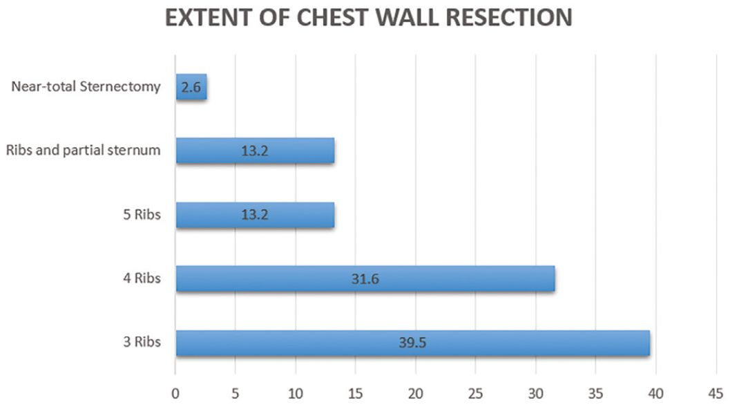
Mean operative time was 185±56 minutes (range, 120–300), and mean blood loss was 295±120 mL (range, 50–550). ICU monitoring was required for a mean of 1±0.5 days. Pain decreased rapidly postoperatively, with mean VAS scores of 4.1±1.1 at 24 hours, 2.0±1.0 at 48 hours, and 1.7±0.7 by postoperative day 7 (). Eleven patients (29%) required no rescue analgesia, while 5 patients (13.2%) required frequent opioid supplementation (). At discharge, 44.7% reported no pain, 34.2% reported mild movement-related discomfort, and 21.1% reported pain at rest.
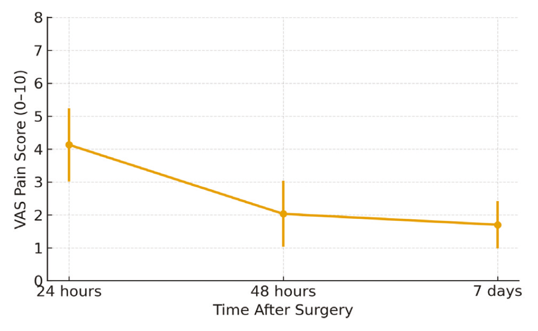
VAS: visual analogue scale, SD: standard deviation, NSAID: nonsteroidal anti-inflammatory drug
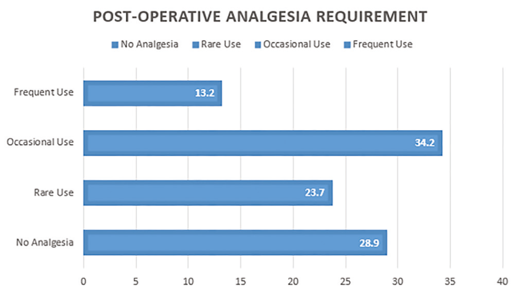
NSAID: nonsteroidal anti-inflammatory drug
Chest wall recoil was preserved in all 38 patients, with no paradoxical movement on clinical examination. At 1 month, 30 patients (79%) had normal chest expansion, 8 (21%) had mild restriction, and 3 (8%) reported dyspnea (). By 3 months, functional outcomes had improved further; only 6% had persistent mild restriction, and no patient reported dyspnea, suggesting a recovery-related rather than a structural limitation.
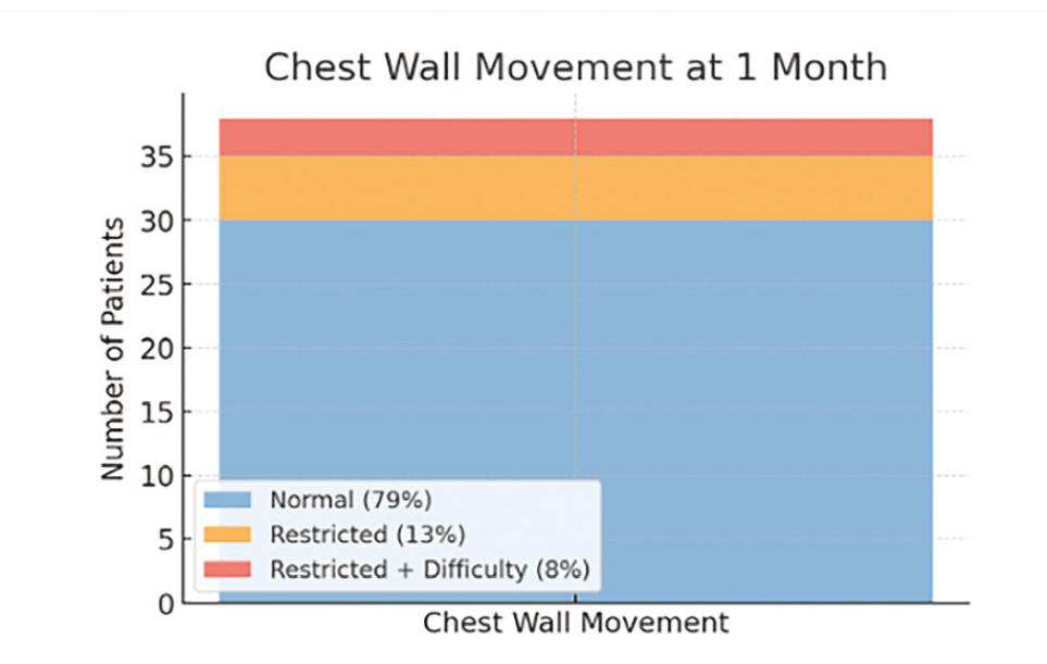
• Normal chest wall movement: symmetrical chest expansion with axillary excursion ≥3 cm or no visible asymmetry compared to the contralateral side
• Restricted movement: visible reduction in expansion (axillary excursion <3 cm or ≥25% less than contralateral side) or patient-reported limitation on deep inspiration
• Intact chest wall recoil: absence of paradoxical inward movement on clinical examination and no flail segment on chest X-ray
Subjective cosmetic deformity was reported by 9 patients (24%), whereas 29 patients (76%) reported no noticeable change from their preoperative appearance. Postoperative complications occurred in 2 patients (5.3%): 1 seroma (2.6%) and 1 case of chronic chest pain lasting >6 weeks (2.6%). No hematoma, wound infection, wire-related failure, implant fracture, or reconstruction instability were observed. R0 margins were obtained in all cases, and local recurrence occurred in one patient during follow-up. No 30-day mortality was recorded.
The mean postoperative hospital stay was 4.18±0.89 days (range, 2–5 days).
DISCUSSION
Over this two-year period, twisted No. 5 stainless steel wire reconstruction demonstrated favorable functional, cosmetic, and perioperative outcomes following oncologic chest wall resection. The technique resulted in low morbidity, no perioperative mortality, and preservation of chest wall physiology in all patients. No paradoxical movement or loss of recoil was observed clinically and on chest X-ray, suggesting that the wire construct provides adequate early biomechanical stability. Although these short-term findings are encouraging, they warrant validation through objective long-term assessments such as pulmonary function testing and dynamic imaging.
The preference for twisted double-stranded wires rather than looped or single-stranded wires was central to our approach. Twisting increases tensile strength, reduces loosening under cyclic respiratory forces, minimizes slippage or rib-edge cut-through, and allows fine contour adjustment. These properties likely contributed to the stable approximation of rib stumps and to the preservation of physiological recoil. Theoretical advantages over non-twisted configurations, such as improved grip and resistance to multidirectional stress, were not biomechanically tested in this study, but were supported by clinical performance. Chest wall reconstruction using stainless steel wires may raise concerns during cardiopulmonary resuscitation or blunt trauma, particularly for defects overlying the cardiac region, as rigid constructs can theoretically alter force transmission and pose a risk of internal injury if not properly secured. With our technique, these risks were minimized by careful contouring and trimming of wires, with twisted ends buried and directed away from the underlying viscera, while the interlocking lattice configuration helped to distribute forces evenly and to preserve some physiological chest wall compliance by avoiding overtightening.
Functional recovery was favorable, with 79% of patients demonstrating normal chest mobility at 1 month and 94% at 3 months. Early mild restriction (21% at 1 month) appeared to reflect postoperative recovery rather than structural instability. By 3 months, dyspnea had resolved entirely and persistent restriction had decreased to 6%, reinforcing the concept that early respiratory limitation is transient. Our outcome assessment, conducted at 1 month, 3 months, and at extended follow-up (to almost 2 years for early cases), revealed no late wire failure, deformity, or respiratory impairment. In our series, no wire fractures, dislocations, or rib-stump cut-throughs were observed, likely due to careful technical strategies, such as avoiding excessive wire tension, using multiple fixation points, favoring circumferential anchorage around rib stumps, and employing a semi-rigid construct that allows limited micro-mobility. Nevertheless, these findings reflect our short-term clinical experience.
Our results meet the criteria for successful semi-rigid reconstruction, achieving stability with preserved mobility. These findings are consistent with a recent overview of 16 studies (n = 1,089) evaluating non-rigid methods, which reported similarly low complication and satisfactory functional outcomes. Cosmetic satisfaction was acceptable, with 9 patients (24%) reporting subjective cosmetic misalignment. Compared with mesh–bone cement composite techniques, known for increased rigidity, infection risk, and operative burden,- our wire-based approach maintained contour while avoiding excessive stiffness.
Operative parameters (mean operative time 185 minutes; blood loss 295 mL) aligned with studies for bone-cement reconstructions, where reported medians reach 240 minutes and 500 mL. Hospital length of stay averaged 4.18 days, and ICU length of stay averaged 1 day. Complications occurred in only 5.3% of patients (seroma and chronic pain); there were no infections, hematomas, wire failures, or reconstruction instability. These rates aligned with rigid systems such as titanium plates or methyl methacrylate “sandwich” constructs, which report complication rates between 10% and 30% and respiratory morbidity., ,
Hybrid techniques combining bone cement with twisted wires for neorib construction have shown promising mechanical strength and cosmetic outcomes,- but introduce cement-related risks including infection, excessive rigidity, and potential for implantation syndrome. Chest wall reconstruction using only stainless steel wires and mesh has been recently reported in India and has results similar to our cohort.
Importantly, the technique is accessible, inexpensive, and particularly advantageous in resource-limited environments. At our center, a pack of eight No. 5 wires costs PKR 3000 (10 USD); typical reconstructions require 5–6 wires, and mesh (20 USD). Despite its simplicity, the technique produced feasible and reproducible outcomes. The Cleveland Clinic’s experience with titanium mesh (n = 22) reported excellent oncologic margins and no respiratory complications, but implant fractures occurred in 68% of patients. The MD Anderson 20-year series (n = 59) reported 62% morbidity and 8.5% mortality, and a recent European study showed 19% morbidity and 4% reconstruction failure following oncological chest wall reconstruction with various prosthesis. Our data show that wire-based reconstruction is an effective, practical, and cost-efficient alternative, as indicated by our early outcomes.
Study Limitations
This study is subject to several limitations inherent in its retrospective design, including the potential for incomplete data capture, missing variables, and reliance on documentation that was not originally intended for research purposes. The study also has a modest sample size, lacks a comparator group, and has heterogeneous follow-up durations, which may limit the generalizability of the findings. Functional outcomes were assessed primarily by clinical evaluation without objective measures such as pulmonary function tests, quality of life scores, or dynamic imaging, thereby introducing observer bias. Visual evaluation of chest wall motion lacks standardized scoring and is subject to interobserver variability. Analgesic consumption could not be converted to morphine milligram equivalents because of the retrospective study design and the heterogeneity of analgesic protocols. Larger prospective studies incorporating standardized functional testing and long-term follow-up are needed to validate these findings and compare wire-based constructs with titanium or bone-cement systems.
CONCLUSION
Twisted stainless steel wiring represents a pragmatic and affordable option for chest wall reconstruction, offering acceptable structural support, preservation of respiratory mechanics, and satisfactory early clinical and cosmetic outcomes in selected patients. However, given that this study is an uncontrolled retrospective case series, our findings should be interpreted as descriptive and hypothesis-generating. Steel wire–based reconstruction should be viewed as an alternative whose relative role requires further clarification through well-designed prospective and comparative studies.
Footnotes
Ethics
Ethics Committee Approval: Institutional Review Board approval was obtained from the Services Institute of Medical Sciences, Services Hospital, Pakistan (decision no.: IRB/2025/1653/SIMS) on 21.08.2025.
Informed Consent: Informed consent was waived by the Institutional Review Board. All patient data were anonymized to protect confidentiality, and the study adhered to the Declaration of Helsinki and institutional ethical standards. Written informed consent was obtained from all patients for the publication of their clinical data and any accompanying images.
Authorship Contributions
Surgical and Medical Practices: M.S.N., Z.S., R.S., Concept: M.S.N., Design: M.S.N., Z.S., H.I., Data Collection or Processing: M.S.N., Z.S., R.S., Analysis or Interpretation: Z.S., H.I., Literature Search: H.I., R.S., Writing: Z.S., H.I., R.S.
Conflict of Interest: No conflict of interest was declared by the authors.
Financial Disclosure: The authors declared that this study received no financial support.
References
Sanna S, Brandolini J, Pardolesi A, et al. Materials and techniques in chest wall reconstruction: a review. J Vis Surg. 2017;3:95.
Khullar OV, Fernandez FG. Prosthetic reconstruction of the chest wall. Thorac Surg Clin. 2017;27(2):201-208.
Dahan M, Brouchet L, Berjaud J, Garcia O. [Surgery of chest wall tumors]. Ann Chir Plast Esthet. 2003;48(2):93-98.
Suzuki K, Park BJ, Adusumilli PS, et al. Chest wall reconstruction using a methyl methacrylate Neo-Rib and Mesh. Ann Thorac Surg. 2015;100(2):744-747.
Gerrah R. An improved chest wall reconstruction technique with twisted steel wire-reinforced neoribs. Ann Thorac Surg Short Rep. 2023;1(1):111-114.
Ram D, Darlong LM, Sharma A, Bansal A, Shukla H. Chest wall reconstruction using steel wire in a case of chondrosarcoma rib: a novel technique of neo-rib. Indian J Thorac Cardiovasc Surg. 2017;33(2):187-189.
Hines CB. Understanding bone cement implantation syndrome. AANA J. 2018;86(6):433-441.
Vanstraelen S, Bains MS, Dycoco J, et al. Biologic versus synthetic prosthesis for chest wall reconstruction: a matched analysis. Eur J Cardiothorac Surg. 2023;64(6):ezad348.
Vanstraelen S, Ali B, Bains MS, et al. The contribution of microvascular free flaps and pedicled flaps to successful chest wall surgery. J Thorac Cardiovasc Surg. 2023;166(4):1262-1272.e2.
Vanstraelen S, Moonsamy P, Bains MS, et al. Biosandwich technique for extensive chest wall reconstruction in patients with complex defects. JTCVS Tech. 2023;18:164-167.
Ng CS. Recent and future developments in chest wall reconstruction. Semin Thorac Cardiovasc Surg. 2015;27(2):234-239.
Iqbal S, Ali S, Fatimi SH. Novel reconstruction of neosternum with steel wires for recurrent chondrosarcoma. Ann Thorac Surg. 2022;113(5):e371-e374.
Colella S, Brandimarte A, Marra R, et al. Chest wall reconstruction in benign and malignant tumors with non-rigid materials: an overview. Front Surg. 2022;9:976463.
Weyant MJ, Bains MS, Venkatraman E, et al. Results of chest wall resection and reconstruction with and without rigid prosthesis. Ann Thorac Surg. 2006;81(1):279-285.
Kilic D, Gungor A, Kavukcu S, et al. Comparison of mersilene mesh-methyl metacrylate sandwich and polytetrafluoroethylene grafts for chest wall reconstruction. J Invest Surg. 2006;19(6):353-360.
Menon VS, Padhy AS, Nittala R. Short-term outcomes of chest wall resections. Indian J Thorac Cardiovasc Surg. 2025;41(9):1173-1179.
Koole D, Bemelman M, Schijen J, de Fijter M, van der Niet J. Reconstruction of chest wall defects using a technique involving mesh, titanium plates, and a pedunculated muscle flap. Korean J Thorac Cardiovasc Surg. 2018;51(5):308-311.
Brascia D, Mangiameli G, Marulli G. Complex chest wall reconstruction after failure: a literature review. J Thorac Dis. 2024;16(7):4780-4793.
Darlong LM, Kushwaha NK, Garg N, et al. Neo-Rib technique of chest wall reconstruction following oncological resection: an institutional experience. Indian J Surg Oncol. 2025;17(7):1582-1594.
Tasnim S, Barron JO, Raja S, Murthy SC, Raymond DP. Five-year experience with titanium mesh for rigid chest wall reconstruction. JTCVS Tech. 2024;28:180-190.
Corkum JP, Garvey PB, Baumann DP, et al. Reconstruction of massive chest wall defects: a 20-year experience. J Plast Reconstr Aesthet Surg. 2020;73(6):1091-1098.
Fernandez Gonzalez AM, Matilla JR, Pipek OA, et al. Long-term outcome and prognostic factors after chest wall resection and reconstruction. Interdiscip Cardiovasc Thorac Surg. 2025;40(6):ivaf126.